X Rays Stack Up
A relatively new x-ray imaging technique has made its first foray into the third dimension. Using a computer algorithm and high energy x rays, researchers were able to visualize two nanometer-scale, etched patterns stacked one on top of the other. The 19 August print issue of PRL describes this step toward atomic-scale probing of irregular structures such as material defects, nanostructures, and perhaps the many protein molecules that resist crystallization.
X-ray microscopy along the lines of light microscopy resolves at best down to 30 nanometers because x rays are difficult to focus. So for many applications researchers turn to diffraction, which requires a crystal, but can achieve resolutions of a few angstroms or better. Crystal diffraction creates a pattern of spots of varying intensities and positions related to the repetitive arrangement of atoms within the sample.
But not everything comes in perfect crystals. Some techniques exist for handling partially crystalline materials, but many other target structures have no periodicity at all, such as material defects and the large number of proteins that refuse to crystallize. Constructing a holographic image from two x-ray beams is one potential option for imaging these holdouts, but there is a way of doing it with one beam. Non-crystalline formations produce weak and continuous diffraction patterns–rather than spots–in which every point contains some information about the original structure. “Oversampling” this pattern with a detector that has many pixels can extract enough information to reconstruct the sample structure. So far physicists have used this new technique to image only two-dimensional slices of 3D objects.
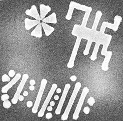
Now Jianwei Miao of Stanford University and his colleagues have condensed a series of these oversampled diffraction patterns into a simple 3D structure for the first time, making use of a powerful x ray source and a computer algorithm they developed. Their sample was a pair of nanoscale patterns–each one included a simulated DNA strand, a circuit pattern, and a star–etched in nickel with an electron beam and sandwiched together. They hit the object at 30 different angles with 6 keV x rays from the SPring-8 synchrotron at the Institute of Chemical and Physical Research (RIKEN) in Japan, then mapped each of the diffraction patterns onto a three-dimensional coordinate system. Information garnered from oversampling gradually builds up a 3D view from an initial random reconstruction of the structure. They resolved the target down to 50 nanometers, but Miao hopes to reach the atomic level eventually, which he says will require longer exposure times, better detectors and greater computing power.
The particular method used here has some drawbacks, says Ian Robinson of the University of Illinois at Urbana-Champaign, who is working on another oversampling technique for solving 3D structures. The calculations are complicated, and taking multiple views at different angles can be imprecise, he says. However, “I certainly agree the oversampling method has great potential.”
Potential applications include imaging disordered materials and biological samples such as cells and viruses. And because the method doesn’t require crystals, Miao explains, it could potentially image single molecules, perhaps in conjunction with a free electron laser–a large facility that provides an x-ray laser beam. “If that’s true, that’s going to revolutionize the structural molecular biology field,” he says. Of course, adds Robinson, such an experiment is probably 20 years down the road.
–JR Minkel
JR Minkel is a freelance science writer in New York City.