Blurring Clarifies Cell Movements
To see how a cell moves and molds its shape, get a low-tech microscope and turn the knob until it’s out of focus, Brazilian researchers advise. In the May Physical Review E they show mathematically that the bright and dark features of a “defocused” image reveal the topography of cell surfaces. With blurry movies, they recorded moving ripples, which their experiments suggest may figure in cells’ strategy for engulfing prey.
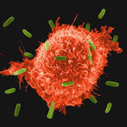
You only need to look at an amoeba to realize that biological cells are no ordinary sacks of goo. Biologists know that cells direct their motion by slowly restructuring the springy scaffolding underneath their outer envelope, or membrane. But many details of the process remain mysterious. Already researchers have brought various high-tech tools to the task, but few of them are easy.
At the Federal University of Minas Gerais in Belo Horizonte, Brazil, Oscar Mesquita had in mind to measure movements in macrophages–scavenging white blood cells, which his medical colleagues hope to induce to swallow drug particles. He peeked through the microscope before focusing and was surprised to see dark ripples that moved inward from the faint periphery of the cell. The dark bands changed to light as he fiddled with the focus. Mesquita says he knew the drifting shadows somehow reflected motions of the cell, but he didn’t know exactly what they meant. He and his colleagues plunged into optical mathematics and emerged with an approximate formula. “What we found is that the contrast of the defocused image is related to the curvature of the object,” he says.
The physical explanation was evident in their equation, which reproduced the “lens maker’s formula” of introductory physics. This means the hills and dales of the cell surface act as convex or concave lenses, Mesquite says, creating contrast by concentrating or diluting the light rays that pass through them.
With their formula, Mesquite and his colleagues were able to convert shades of gray to curvatures, from which they produced 3D pictures of the ripples. They also clocked the ripples’ speeds. When the researchers treated macrophages with a drug that weakens the understructure of the membrane, the ripples disappeared. At the same time, the cells took more than 40 times longer to swallow a yeast pellet, suggesting that the ripples may be associated with the swallowing process.
Researchers are impressed with what the Brazilian team was able to do with such simple technology. Their method is “within the capacity of any cell biology microscope,” says Washington University’s Elliot Elson. He says the ripples are the spitting image of undulations he and colleagues reconstructed 15 years ago using slices through many macrophages, “a much more cumbersome approach.”
Erich Sackmann of the Technical University of Munich (TUM) is less impressed, since the technique doesn’t have high resolution. “I doubt somewhat whether the technique yields useful quantitative information” regarding physical properties such as membrane stiffness, he says. But his TUM colleague Andreas Bausch thinks the ability to capture features in motion could be important. “I think there are all kinds of potential applications here,” he says.
–Oliver Baker
Oliver Baker is a freelance science writer based in Davis, CA.