Tips for Better Fluorescence

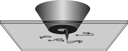
Two improved fluorescence microscopes, reported in the 29 October and 12 November issues of PRL, could allow researchers to see individual protein molecules on the surface of a living cell. Both teams of researchers obtained fluorescence images by dipping a needle-like “tip” into the focus of the laser used to create the fluorescence. One team improved the positioning of their tip, while the other channeled the laser light through a narrow aperture before letting it hit the tip.
Optical fluorescence microscopy, in which a laser at one wavelength stimulates a sample to fluoresce at a different wavelength, should be a good way to view proteins without disrupting them. “But the glaring drawback of normal fluorescence microscopy is the resolution,” one-half the wavelength of the laser light, says Jordan Gerton of the University of Utah. That means 250 to 300 nanometers, whereas the average protein is 1 to 10 nanometers across, he says.
To break this resolution limit, researchers use the extremely sharp point or tip from an atomic force microscope (AFM) to enhance the fluorescence signal–one form of a so-called scanning near-field optical microscope (SNOM). The tip is placed at the focal point of a laser beam–just above the sample surface–and the beam and tip move across the sample in tandem. This technique increases the electric field and therefore the fluorescence at the AFM tip because charges tend to concentrate at sharp corners, such as the end of a lightning rod. Although this method resolves images at close to 10 nanometers, it is plagued by poor signal quality because metal tips can “quench,” or siphon excited electrons away from fluorescent dyes, preventing photon emission.
Gerton, team leader Stephen Quake of the California Institute of Technology in Pasadena, and their colleagues took precise fluorescence measurements that showed that the tip would have to touch the sample to get the best fluorescence and maximum resolution, whereas others had let the tip hover a few nanometers above the sample for convenience. Touching the sample increased its fluorescence up to 20-fold beyond the background signal from the laser alone, giving them four times the contrast of previous efforts. They used a silicon tip to avoid quenching, even though metal can theoretically create more fluorescence. By scanning it over their sample–a 5-nanometer-wide semiconductor nanocrystal sitting on a glass surface–the team was able to observe high quality fluorescence with at least 10-nanometer resolution.
An alternative to an AFM-type device is to shine a laser through a narrow glass fiber onto a sample, which reduces background fluorescence. Such microscopes usually have higher sensitivity to faint fluorescence but lower spatial resolution than tip-based microscopes. Heinrich Frey, Reinhard Guckenberger, and colleagues at the Max Planck Institute for Biochemistry in Martinsried, Germany, combined sensitivity and resolution by putting a metal-coated tip on the rim of a 100-nanometer-wide hole in a tapered glass fiber. They shined a laser down the fiber to illuminate the tip and their sample, a mica surface covered with DNA molecules whose ends were labeled with fluorescent dye. Although the team did have to contend with quenching, their low background signal allowed them to resolve individual dye molecules separated by as little as 10 nanometers. Both groups say their methods should be able to achieve even finer resolution.
“The new results stand out because of their quality,” says Lukas Novotny of the University of Rochester in New York. “They clearly demonstrate that tip-enhanced microscopy is possible on the single molecule level” without significant quenching. That could point the way to resolving, mapping, or even manipulating single proteins on cell surfaces, he says.
–JR Minkel
JR Minkel is a freelance science writer in New York City.


