Ghost Imaging with Electrons
The expression “ghost imaging” might conjure up blurry photos of ghoulish visitors, but it’s actually a sophisticated approach to imaging objects that may not be suited to conventional imaging methods. To date, ghost imaging has employed visible light, x rays, and even helium atoms as illumination sources. And now, researchers have demonstrated a ghost-imaging setup for electrons. Compared to standard electron imaging, this approach can reduce the acquisition time and the radiation dose on the sample—an asset for samples easily damaged by radiation, like biomolecules.
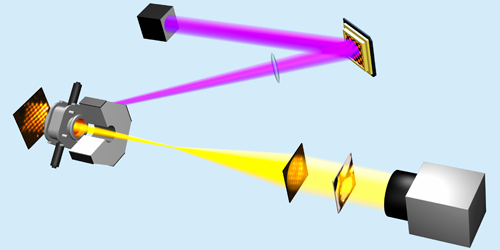
Conventional ghost imaging constructs images of objects via correlations between the spatial profile of a “reference” beam and the intensity of a “signal” beam. The signal beam reflects off the target or is transmitted through it and then hits a single-pixel detector. Crucially, the signal beam can contain far fewer photons than the reference beam, avoiding sample damage.
Typically, the reference and signal beams are made by splitting one beam of light. The challenge for electron ghost imaging is that there are no devices that can split a beam of electrons with the typical energies used in imaging experiments. So Siqi Li of the SLAC National Accelerator Laboratory, California, and colleagues employed a system that replaced the reference beam with a computed version of it, obtained digitally from the incident electron beam. The pattern from this computed reference beam was then correlated with the intensity measured by the single-pixel detector. Using this setup, the team successfully reconstructed the image of a metal ring placed in the path of the signal beam. The authors suggest that similar approaches could be used with other illumination types, potentially extending ghost imaging to ions, plasmas, and neutrons.
This research is published in Physical Review Letters.
–Christopher Crockett
Christopher Crockett is a freelance writer based in Arlington, Virginia.