Reversing Light Scattering with a Handful of Photons
It’s tricky to see inside a turbid medium like biological tissue using visible light, as most of the illuminating photons will get randomly scattered. Now researchers have pushed the limits of a previously studied technique for reversing the effects of strong scattering and shown that it works even when relatively few photons are detected. The team sent a light beam through a thin but highly scattering material and showed that they could reproduce an image of the original beam, even though they only detected about 1000 of the scattered photons. They found that, in theory, there is no lower limit on the number of photons that could lead to useful imaging.
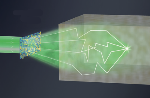
Optical phase conjugation (OPC) is a decades-old technique for reversing the effects of a nearly opaque medium, and it involves two steps. In one version of the method used for imaging, the “recording” step starts with a probe beam sent through the sample to be imaged, with one small point in the sample specially “labeled” by a so-called guide star. For example, one labeling technique uses a focused ultrasound beam to frequency-shift the light that propagates through the guide star point. The guide-star-influenced light that emerges from the sample then interferes with a second light source called the reference. The interference produces a quasirandom array of bright and dark patches—a hologram that contains information about the scattering paths of the light in the sample.
In the “playback” step, another beam goes through the sample in the opposite direction after first passing through a filter that is imprinted with the hologram. This filtering causes the playback beam to undergo the exact reverse of all of the complicated scattering experienced by the original probe light, so that it focuses to a bright spot at the guide star location. This bright light is enough to image that region, for example by conventional fluorescence microscopy. To produce a complete 3D image of a sample, the guide star is moved around from point to point, and in principle the technique could probe several millimeters into a mouse brain, for example.
The trouble is, in highly turbid media, not much light emerges during the recording step. For biological samples, moreover, one can’t compensate by collecting data for a long time, because things are constantly moving. So researchers have assumed that deep-tissue imaging wouldn’t be possible with OPC. However, optical physicists Mooseok Jang and Changhuei Yang at the California Institute of Technology in Pasadena, collaborating with biomedical imaging specialist Ivo Vellekoop of the University of Twente in the Netherlands, have now shown that OPC can work for signals previously thought much too weak to be useful.
The researchers used a very weak probe beam to test the intensity limits of the approach. For their experiments they used a 0.45-mm-thick layer of synthetic opal, a highly scattering material. The team sent their probe beam through the opal and created an interference pattern between the resulting scattered light and a reference beam, to produce the hologram for the playback beam. When the playback beam was shone back through the opal and hit a detector, it recreated an image of the probe beam, the equivalent of lighting up the guide star location.
Jang and colleagues could produce such an image even when the scattered probe beam contained only about 1000 photons. This is so weak that there were far fewer scattered photons than detector pixels recording the interference pattern, perhaps as few as 0.004 photons per pixel. Paradoxically, however, these photons make their presence felt throughout the entire interference pattern, so that every detector pixel records information needed for making the phase-conjugated playback beam.
“Phase conjugation-based approaches currently provide light focusing capability only for tissue thicknesses of no more than 5 mm or so,” says Jang. “Our study implies that we can significantly extend this range,” because so few photons are required.
It’s not just imaging that might benefit, adds Vellekoop. Combined with the technique of optogenetics, in which genetic and thus cell activity can be controlled by light, it might be possible to focus light and thus optically control the activity of individual neurons, deep within brain tissue.
“This is convincing work with results that could affect applications,” says Yaron Silberberg, an optical physicist at the Weizmann Institute of Science in Rehovot, Israel. He says the research shows that “even when there are fewer photons than there are resolution elements in the phase-conjugating setup, the technique can still be used effectively.”
This research is published in Physical Review Letters.
–Philip Ball
Philip Ball is a freelance science writer in London. His latest book is How Life Works (Picador, 2024).


