Electrons Film Phonon Dynamics in Full
Atoms in solids are always in motion, vibrating about some average position. At a given temperature, the manner in which they jiggle depends on the available vibrational states—quantized modes called phonons [1]—and on how such states are populated. This simple picture does a good job of describing equilibrium states, but it leaves out an enormous amount of detail about how vibrational processes unfold in real time. On timescales as short as femtoseconds (fs), electrons can transfer energy to vibrations, and energy gets shuffled between phonons that are coupled to each other. These ultrafast processes play critical roles in many complex phenomena, including phase transitions, superconductivity, and other forms of emergent behavior. To date, unfortunately, our understanding of phonon dynamics relies mostly on theory, because of the difficulty of probing collective vibrational phenomena [2]. An experimental method capable of providing a full picture of phonon dynamics would be immensely useful. Now, a team led by Bradley Siwick at McGill University in Canada has demonstrated that extremely short packets of electrons can measure the dynamics of all the phonon modes of a material. After photoexciting electrons in a graphite sample, their experiments track how the energy from this excitation is transferred from electrons to phonons and then redistributed among specific phonon modes [3].
Phonons have effects on all manner of solid-state properties. For instance, they govern the thermal conductivity of semiconductors and affect the electrical resistivity of metals. A detailed picture of phonon behavior is thus essential for understanding many thermal, electrical, and optical properties of materials. Static experimental methods like infrared and Raman spectroscopy, x-ray diffuse scattering, and neutron scattering have provided a wealth of information by measuring phonons in the frequency domain. New tools continue to emerge, such as high-energy-resolution transmission electron microscopes [4], which pave the way to vibrational spectroscopy with atomic-scale resolution. These methods are all based on scattering measurements. They extract phonon frequencies and amplitudes by monitoring the change in energy and/or momentum of incoming particles (photons, electrons, or neutrons) as they scatter from atoms or electrons in a sample, exciting phonons in the process.
These approaches deliver static measurements of average phonon properties. A different strategy, however, is needed to monitor phonons on their natural timescales: femtoseconds to picoseconds (ps). The advent of short-pulsed lasers provided a way to extend static methods into the ultrafast time domain. Several time-resolved methods for studying phonon dynamics have been demonstrated [5–7]. They generally operate via a so-called pump-probe approach: a sample is first excited (pumped) with a laser pulse and then probed with a second laser pulse some time after excitation. The evolution of various spectroscopic signals—such as Raman shifts or changes in vibrational absorption lines—can reveal phonon dynamics, with a temporal resolution limited by the pulse durations and a spatial resolution limited by the laser wavelength. These methods, however, are typically limited to specific aspects of phonon dynamics. For instance, they might be able to probe phonons only at certain frequencies or of a given type (acoustical or optical phonons).
Siwick’s team uses femtosecond packets of energetic (100-keV) electrons as their probe, rather than photon pulses. This provides a direct probe of transient atomic positions with a spatial resolution far exceeding that of optical methods. Using brief packets of electrons in ultrafast diffraction and microscopy experiments is not new [8–10]. Previous studies accessed phonon dynamics by monitoring Bragg reflections—which can be modulated by phonon oscillations—or by acquiring real-space images of coherent lattice motions. While powerful, such approaches often provide only partial information on a subset of phonons. The team’s time-resolved method exploits the fact that in an electron scattering experiment, structural information is not only contained in electrons coherently scattered into a pattern of discrete Bragg spots but also in those electrons that are scattered with a broad range of momenta into a “diffuse” scattering signal. Accordingly, the team dubbed their approach ultrafast electron diffuse scattering (UEDS).
Unlike Bragg reflections, diffuse scattering arises from deviations in the lattice periodicity. Phonons can contribute to diffuse scattering because they cause fluctuations of the atoms’ positions in the lattice. Previous static studies showed that the diffuse scattering contains information on specific phonons [11]. Remarkably, the authors’ UEDS setup can perform similar measurements with femtosecond time resolution. To simplify the analysis of the UEDS signal, the team employed a thin film of graphite—a highly symmetric material made of identical 2D carbon layers. They showed that, because of selection rules imposed by symmetry, the dynamics of specific phonons can be extracted by analyzing particular points in the diffuse scattering pattern.
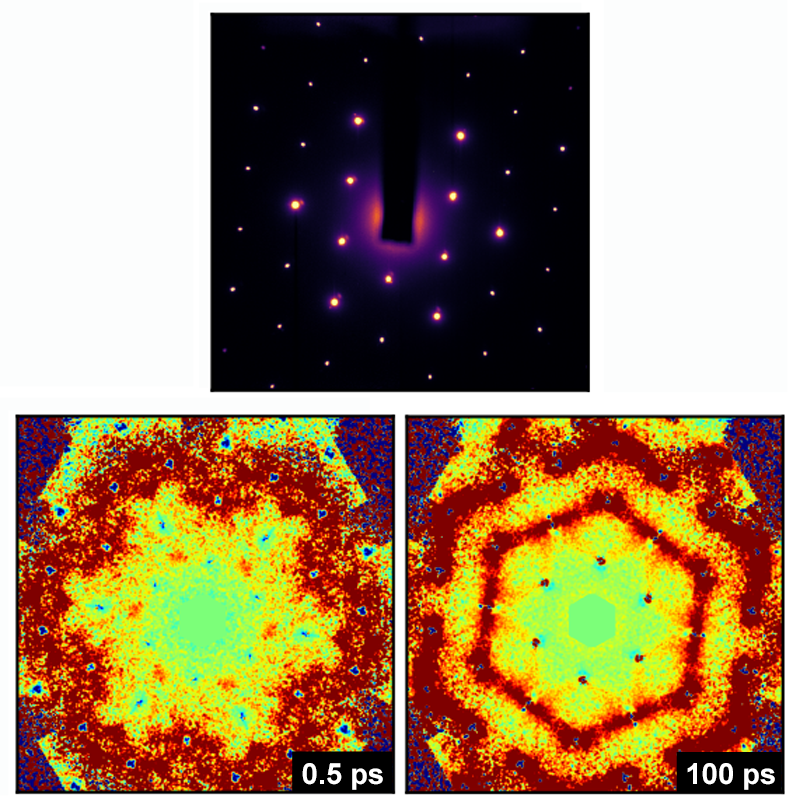
The researchers pumped a thin graphite film with a 35-fs near-infrared laser pulse, which created a population of excited charge carriers in the material. They then probed the sample with ∼ 100-fs electron packets. The angular distribution of the diffusely scattered electrons displayed symmetric patterns, whose shapes directly revealed the population, coupling, and decay of the optical and acoustic phonon modes of the material (Fig. 1). The team acquired a series of scattering patterns at time intervals of about 200 fs. By analyzing these snapshots, they obtained a clear and detailed picture of the underlying dynamics of all of graphite’s phonon modes—optical and acoustic as well as transverse and longitudinal. Such dynamics proceed as follows: The photoexcited charge carriers quickly thermalize via carrier-carrier scattering. Soon thereafter, electron-phonon coupling populates several optical phonon modes that, in turn, transfer their energy to acoustic phonons. On longer timescales, the system finally relaxes back to the ground-state distribution of phonons at thermal equilibrium. Siwick and his colleagues were able to selectively map the entire set of transient coupling processes between electrons and phonons and between different phonons, and they were able to deduce the strengths with which specific phonon modes couple to each other.
The UEDS method will be a powerful tool for studying complex phonon dynamics in solids, which might open new and exciting research directions. If the technique can be extended to crystals having less symmetry than graphite, it might be applied, for example, to low-symmetry molecular crystals that hold promise for organic-semiconductor applications. Understanding electron-phonon and phonon-phonon coupling will be key to devising ways to control electron transport in these materials. UEDS studies may also help researchers probe the relationship between phonons and superconductivity or decipher how electron-phonon coupling contributes to spectacular phenomena like colossal magnetoresistance in strongly correlated electron materials.
This research is published in Physical Review B.
References
- M. Born and K. Huang, Dynamical Theory of Crystal Lattices (Oxford University Press, Oxford, 1954)[Amazon][WorldCat].
- S. Baroni, S. de Gironcoli, A. Dal Corso, and P. Giannozzi, “Phonons and Related Crystal Properties from Density-Functional Perturbation Theory,” Rev. Mod. Phys. 73, 515 (2001).
- M. J. Stern, L. P. René de Cotret, M. R. Otto, R. P. Chatelain, J.-P. Boisvert, M. Sutton, and B. J. Siwick, “Mapping Momentum-Dependent Electron-Phonon Coupling and Nonequilibrium Phonon Dynamics with Ultrafast Electron Diffuse Scattering,” Phys. Rev. B 97, 165416 (2018).
- O. L. Krivanek et al., “Vibrational Spectroscopy in the Electron Microscope,” Nature 514, 209 (2014).
- T. Henighan et al., “Generation Mechanism of Terahertz Coherent Acoustic Phonons in Fe,” Phys. Rev. B 93, 220301(R) (2016).
- J. Koivistoinen, P. Myllyperkiö, and M. Pettersson, “Time-Resolved Coherent Anti-Stokes Raman Scattering of Graphene: Dephasing Dynamics of Optical Phonon,” J. Phys. Chem. Lett. 8, 4108 (2017).
- Y.-H. Cheng, F. Y. Gao, S. W. Teitelbaum, and K. A. Nelson, “Coherent Control of Optical Phonons in Bismuth,” Phys. Rev. B 96, 134302 (2017).
- L. Waldecker, R. Bertoni, H. Hübener, T. Brumme, T. Vasileiadis, D. Zahn, A. Rubio, and R. Ernstorfer, “Momentum-Resolved View of Electron-Phonon Coupling in Multilayer WSe2,” Phys. Rev. Lett. 119, 036803 (2017).
- T. Konstantinova et al., “Nonequilibrium Electron and Lattice Dynamics of Strongly Correlated Bi2Sr2CaCu2O8+𝛿 Single Crystals,” Sci. Adv. 4, eaap7427 (2018).
- D. R. Cremons, D. X. Du, and D. J. Flannigan, “Picosecond Phase-Velocity Dispersion of Hypersonic Phonons Imaged with Ultrafast Electron Microscopy,” Phys. Rev. Mater. 1, 073801 (2017).
- M. Holt, Z. Wu, H. Hong, P. Zschack, P. Jemian, J. Tischler, H. Chen, and T.-C. Chiang, “Determination of Phonon Dispersions from X-Ray Transmission Scattering: The Example of Silicon,” Phys. Rev. Lett. 83, 3317 (1999).