Bringing High-Resolution X-Ray Imaging to the Laboratory
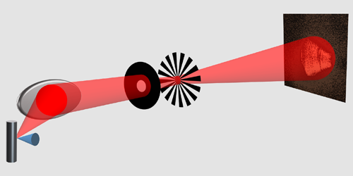
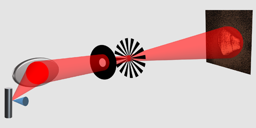
X-ray ptychography is a powerful new technique that uses the quantitative phase information in transmitted radiation to create nanometer-resolution images. Researchers have applied the method to image everything from biological tissues to archaeological artifacts to semiconductor nanostructures, but until now, the intense and coherent x-ray sources that it requires have been available only at expensive synchrotron or free-electron-laser facilities. Darren Batey, of the Diamond Light Source in the UK, and colleagues hope to change that with their demonstration of a ptychography technique that uses a more accessible, laboratory-scale x-ray source [1].
Like conventional x-ray ptychography, the team’s method involves scanning an object with a coherent x-ray beam and reconstructing an image from the resulting interference patterns. The quality of the image depends on the amount of coherent x rays, so Batey and colleagues use the most powerful laboratory x-ray source available. This source produces x rays by slamming electrons into a liquid gallium target, tolerating electron currents that would melt the solid metal target in a standard laboratory-scale x-ray source. Even so, this source is still much dimmer than synchrotrons and free-electron lasers, and its output spans a larger range of frequencies.
To make up for the source’s large frequency range, the researchers use a photon-counting detector containing a spectrometer that narrows the radiation bandwidth after collection. To compensate for its relative lack of intensity, they use a reconstruction algorithm that is robust to beam instabilities that may occur during the necessarily long acquisition times. The researchers say that by integrating these innovations, it is now feasible to deliver the analytical power of ptychographic x-ray imaging directly into universities and hospitals, making it a routine tool for research, education, and medical diagnostics .
–Sophia Chen
Sophia Chen is a freelance science writer based in Columbus, Ohio.
References
- D. J. Batey et al., “X-ray ptychography with a laboratory source,” Phys. Rev. Lett. 126, 193902 (2021).


