Strain-Induced Jamming in an Aquatic Cell
In a fraction of the time that it takes to blink, a single-celled organism known as Spirostomum ambiguum can shrink its length by more than half. This process generates twice the g-force of that experienced by a Formula One driver during cornering. This force is also more than 10,000 times greater than that experienced by any other contracting cell. Now Manu Prakash and Ray Chang, both of Stanford University, have a possible explanation for how Spirostomum ambiguum dissipates the large amount of energy produced during its rapid contraction without destroying its internal structure [1]. They say that their finding could inform the design of future shock-absorbing materials.
Resembling tiny worms, Spirostomum ambiguum live in the sediment beds of brackish water habitats where they feast on bacteria. The insides of these organisms—which range in length from 1 to 4 mm—contain large numbers of fluid-filled sacs called vacuoles, an uncommon feature for either single-cell organisms or for cells in general.
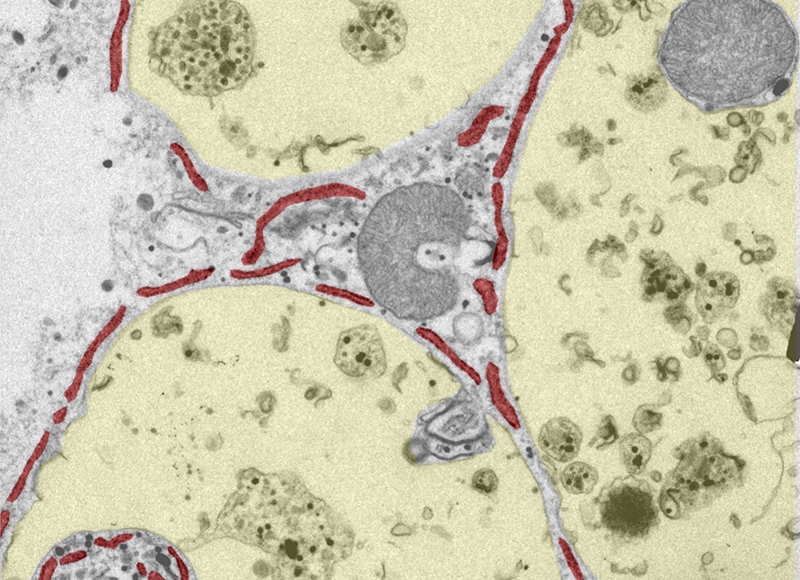
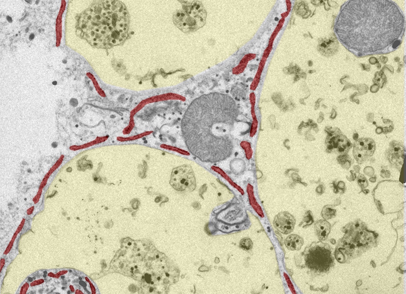
Imaging the insides of Spirostomum ambiguum cells before and after they had undergone a contraction event, Prakash and Chang noticed that these vacuoles appear to be enmeshed in the folds of a membrane-like structure, which they identified as an endoplasmic reticulum. Endoplasmic reticula exist in other organisms and cells, where they produce the proteins needed for cellular functions. But that they can entangle vacuoles in Spirostomum is a previously undocumented finding.
To understand how this entangled membrane–vacuole structure might influence the mechanical properties of Spirostomum ambiguum cells, Prakash and Chang performed two-dimensional simulations of idealized rectangular cells filled with deformable polygons (the vacuoles) and flexible strings (the endoplasmic reticulum). They used values obtained from measurements of living organisms for the density, bending, and stretching properties of the various elements in the cell. They applied a contractile force to the cell and then monitored the positions and shapes of its internal components. The researchers repeated the simulations for cells containing different numbers of strings.
When no strings were present, Prakash and Chang found that the insides of their model cell behaved like a liquid. During contraction, the vacuoles flowed past one another with relative ease and many of the vacuoles swapped positions, a behavior that can disrupt cellular functions in living organisms. The model cells had a higher peak contraction speed than the real cells, and they maintained this speed for several milliseconds. As a result, at the end of the simulation the model cell had shrunk to half the length of the real Spirostomum ambiguum.
Adding in strings, which in the simulation was done by connecting different vacuoles, the researchers observed that the number of vacuole rearrangements during contraction dropped as the number of strings increased. This drop was accompanied by an increase in the effective viscosity of a cell and in the ability of a cell to absorb energy from the contraction. Measuring the number of strings connected to each vacuole as a fraction of the total number of neighboring vacuoles, Prakash and Chang found that for a value of 0.268, the entanglement of the vacuoles and the strings was sufficient to effectively halt vacuole rearrangements. Instead, as the cell shrank the system transitioned from fluid-like to solid-like, with the vacuoles deforming under the contractive forces and jamming each other in place. Under these conditions, the behavior predicted by the simulations matched that measured for living Spirostomum ambiguum cells.
Jamming occurs in systems ranging from sand piles to biological tissues and typically happens when the particle density becomes high enough that the particles can no longer move. Materials that undergo strain-induced jamming—where deformation of a system induces jamming—could be useful for making shock absorbers. To that end, Prakash and Chang are currently developing artificial versions of the entangled Spirostomum ambiguum system that have the right strain-induced jamming properties for such applications.
The results show the importance of internal membrane-like structures in understanding the mechanical response of cells, says Guillaume Charras, a biophysicist at University College London who studies cells and tissues. “That is generally unappreciated and has been overlooked.” For Philip LeDuc, who studies problems at the intersection of mechanical engineering and biology at Carnegie Mellon University in Pennsylvania, the results have broader implications. “The insights here will allow the field to think about many future intersections between biology, physics, and engineering among many disciplines,” he says.
–Katherine Skipper
Katherine Skipper is a science writer based in Bristol, UK.
References
- R. Chang and M. Prakash, “Topological damping in an ultrafast giant cell,” Proc. Natl. Acad. Sci. 120 (2023).


