Modeling Tissue Mechanics with Molten Glass
For thousands of years, humans have crafted glass into everything from useful household items to elaborate story-depicting windows. Researchers at the University of Miami (UM) now add to this extensive repertoire by using glass to study tissue mechanics. The collaboration, which includes scientists and artists, creates glass replications of simple animal tissues and then subjects these glass models to mechanical forces using heat and gravity. At the APS March Meeting, the group presented molten creations that mimic the tissue-stretching patterns observed in the microscopic marine animal Trichoplax adhaerens.
UM biophysicist Vivek Prakash studies fluid flows in animals—and not just the water that flows around marine invertebrates. He also studies the flow of cells within animal tissues. Sheet-like epithelial tissues are made up of collections of connected cells that can constantly move relative to each other when subjected to forces, sometimes jamming together and sometimes rearranging fluidly. Not quite liquid and not quite solid, these tissues stretch, break, and heal in interesting ways. So does molten glass, as Prakash learned a few years ago at a glass art demonstration at UM’s Lowe Art Museum.
That similarity between glass and tissue became the basis of a science-meets-art project in Prakash’s lab. It started with Carolyn Delli-Santi, who began working in the lab as an undergraduate in 2022 studying sea star larvae. These organisms create a “beautiful array of water vortices around themselves to move and feed,” says Delli-Santi. After sketching larvae in their different stages of development, Delli-Santi had the idea of casting these tiny specimens into glass. “I was a science student, and I picked up glass as a minor,” Delli-Santi says. “When I had the opportunity to make this into a serious combination, it was the most exciting thing in the world for me.”
Delli-Santi worked with glass artist Jenna Efrein to fabricate several glass models of sea star larvae in UM’s glass studio. With their flat shape, the sea star pieces can be mounted on a wall as a science-inspired decoration, the artists say. “I have taken the forms, patterns, and physical states of the animals from these projects into my own work,” says Efrein. Their means of movement and physical properties lend themselves well to her work’s conceptual aspects.
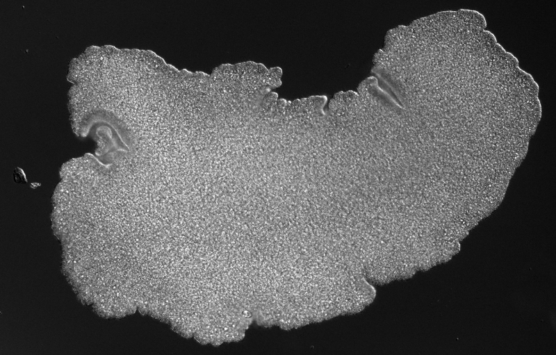
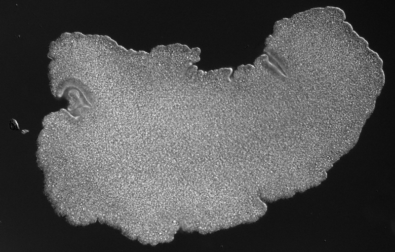
Following the sea star success, Delli-Santi and Efrein teamed up with Prakash to study his research interest, Trichoplax. This simple, 4-mm-wide, multicellular animal lacks muscles or organs. Its flat, amorphous-shaped body consists entirely of two epithelial tissue layers, covered in hair-like cilia, that sandwich a fibrous cavity of fiber cells. Normally, the creature is ductile, changing shape as it crawls along its seafloor environment. But its tissues cross over to being brittle when experiencing mechanical strain [1]. That brittleness can lead to fractures in its epithelium—perhaps as part of its reproductive mechanism—but these fractures can also heal themselves. Fascinated by this shape-shifting creature, Prakash and his colleagues wanted to see if they could recreate its bizarre behavior in a macroscopic analog.
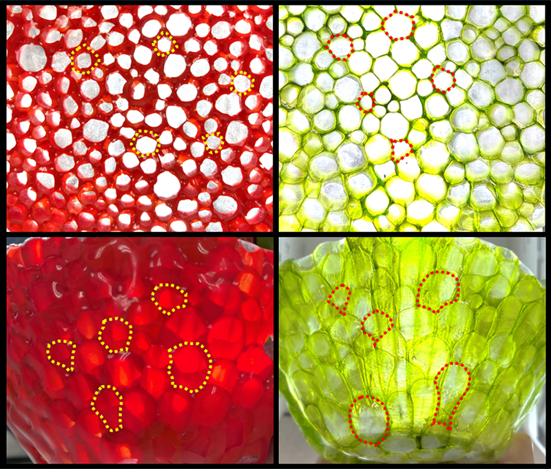
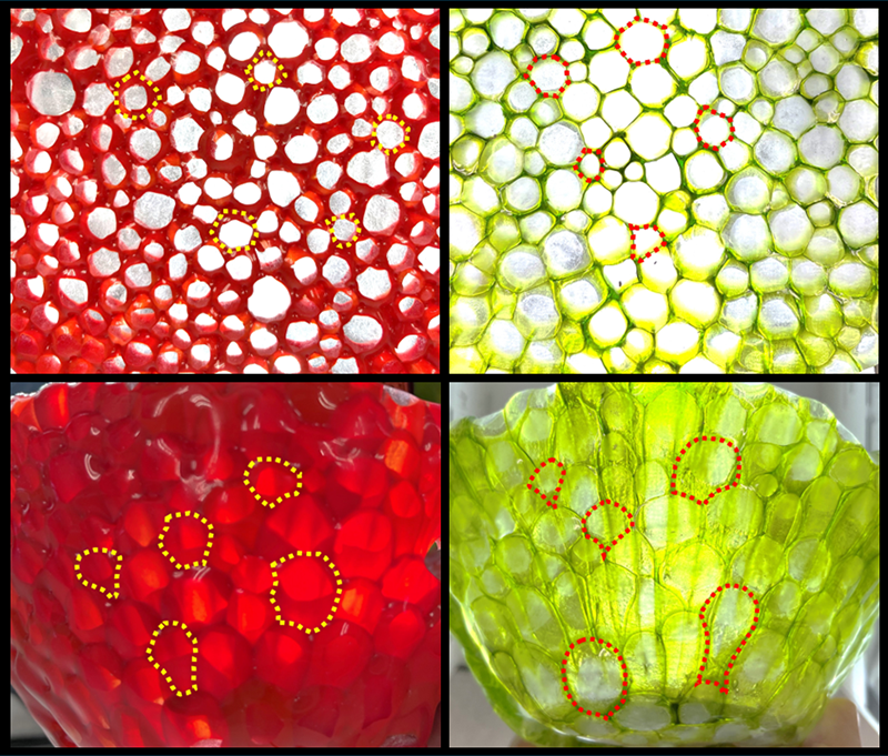
In the glass studio turned biophysics laboratory, the team fabricated Trichoplax models one cell at a time. They first heated molten glass and pulled it into cylindrical shapes that were then cut into 1-cm-high segments to represent the individual cells. A few hundred of these cells were then arranged within a 7-inch-diameter mold and fused into a disk shape. The resulting Trichoplax-like structure was ready to act as a test bed for tissue mechanics [2].
The researchers heated the disk-shaped “tissue” in a kiln to 700 °C, hotter than the transition temperature where glass becomes molten. They then placed the disk on a frustum—a blunted-cone-shaped solid—subjecting it to a gravitational force that pulled on it radially. The molten cells stretched or flowed under the weight of the rest of the tissue, exhibiting a ductile deformation. After this step, the team “froze” the entire glass tissue by quickly cooling it to the annealing point at which movement stops. By freezing the glass dynamics, the researchers obtained a “snapshot” of the deformation at a specific moment in time. “We see that the glass tissue that’s not directly in contact with the center region of the frustum slumps downward as it melts due to heating,” says Gopika Madhu, a physics graduate student working on the project.
The researchers have quantified the changes in cell area and eccentricity as the cells go through their ductile deformations in the glass tissue. “We believe this process has an indirect analogy to the ductile stretching of cells in the tissues of the Trichoplax, which have been shown to be driven by mechanical forces,” says Prakash. The observations reveal an increase in the average cell area during stretching, which confirms mechanical modeling expectations that the cells experience strain under loading. The results also show that the cell eccentricities decrease as a function of the stretch, meaning that they go from an initial circular shape to an elongated elliptical shape—a similar deformation process to that observed in the tissues of Trichoplax. “I had never imagined that you could draw parallels between glass and tissue, so finding this new avenue of research was very exciting,” says Madhu.
Next, the team intends to do computer simulations to obtain stress and strain maps at different locations on the stretched tissue. Limited by the high temperatures, the current dataset only considers before and after stretching; visualizing intermediate points would allow for complete depictions of the ductile and brittle properties—and of other tissue mechanics as a whole—of Trichoplax. “We’re getting data in an artist’s glass shop and turning it into a scientific result,” says Prakash.
–Rachel Berkowitz
Rachel Berkowitz is a Corresponding Editor for Physics Magazine based in Vancouver, Canada.
References
- V. N. Prakash et al., “Motility-induced fracture reveals a ductile-to-brittle crossover in a simple animal’s epithelia,” Nat. Phys. 17, 504 (2021).
- J. Efrein et al., “Glass, Marine Biology, and Physics,” Glass Art Soc. J. 49 (2023), https://issuu.com/glassartsociety/docs/2023_gas_journal.


