Know when to fold ‘em
The complex geometrical shapes of living organisms often emerge from simple mechanical rules. For example, thin sheets of tissue can spontaneously buckle to relieve compressive stress, creating undulating patterns like fingerprints or tree bark.
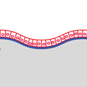
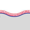
In Physical Review Letters, Edouard Hannezo and colleagues from the Institut Curie in Paris use a buckling model to describe the corrugated inner lining of the intestine, which has a large surface area for absorbing nutrients. In their model, when cells in the single layer at the surface push against each other, they create undulations with wavelengths similar to those seen in animals. But in different parts of the intestine, these undulations ultimately take different forms: In the small intestine, fingerlike “villi” protrude from the surface, while in the colon, cavelike “crypts” extend into the tissue.
The researchers attribute the different morphologies to a new ingredient: variations in the birth and death rate of cells. The division of special cells, which lie deep in the corrugations, continually replenish intestinal cells. The new cells migrate up along the surface to local peaks, where they die. In the model, this migration is impeded by friction from the underlying membrane, so the regions of proliferating cells are more compressed. If the excess compression is big enough, it changes the steady-state shape. In numerical analysis, Hannezo et al. find that this kind of model predicts villi, crypts, or ridges for different choices of parameters. – Don Monroe


