Endoscopy Slims Down
Endoscopes are long, thin tubes that can transmit light deeply into and out of biological tissue and are indispensable for modern surgery and medical diagnostics. Thin endoscopes can be as little as a few millimeters in diameter, but even these scopes can be destructive in certain medical procedures and they have limited imaging resolution. A route to making endoscopes much thinner may emerge from research published in Physical Review Letters. Youngwoon Choi, at Korea University in Seoul, and colleagues have shown they can reconstruct a real image from the scrambled image transmitted by multiple modes of light propagating down a single, few-hundred-micron-thick optical fiber [1]. Their reconstruction relies on the theory for how light propagates in a disordered medium and would allow the multiple optical fibers that currently make up some endoscopes to be replaced by a much narrower, single fiber.
Imaging the insides of biological tissue is notoriously difficult. For one, tissue is highly inhomogeneous, such that most light scatters as it propagates through it. A small fraction of light from a coherent source can penetrate a few hundred microns into tissue, enabling imaging at shallow tissue depths with techniques such as optical coherence tomography, multiphoton microscopy, or confocal microscopy. In contrast, optical tomography techniques that utilize diffusely scattered light can penetrate deeper into tissue, but these techniques aren’t able to resolve features smaller than a few millimeters or centimeters in size [2].
So far, the solution to obtaining high-resolution, microscopic images in deep tissue has been endoscopy: in essence, light is sent down a thin tube that is inserted inside the body, and a series of optical elements relay the image at the end of the endoscope to the outside world. Since the 1960s, many endoscopes have been made out of dense bundles of optical fibers. Multiple-fiber endoscopes (fiberscopes) can contain up to 100,000 individual fibers—each capable of carrying an image pixel—packed into a 1.5-mm-diameter bundle.
In principle, endoscopes could be much thinner if, instead of using multiple fibers in a bundle, it was possible to take advantage of the multiple distinct modes of light that can travel down a single fiber. (These modes are analogous to the different standing wave patterns that form on the surface of a drum.) Unfortunately, when multiple modes travel down a fiber, they can distort and mix in an unpredictable way, scrambling an image. Trying to use multimode fibers in an endoscope has therefore seemed like an impossible task.
Luckily, an intriguing solution for how to send imaging information through a multimode fiber has emerged from the study of how light travels through a disordered medium. If a coherent laser beam is focused at the core of a multimode fiber, it is possible to see, on the other end of the fiber, a complex interference pattern called speckle. This speckle comes from the multiple and random reflections of light as it propagates along the fiber, and it is similar to the speckle pattern that arises when coherent light propagates through a scattering medium, such as the fat globules in a glass of milk. Indeed the two phenomena are related: they come from the fact that a multimode fiber and a scattering system are both complex linear systems that mix, in a complex way, the light that impinges on them. Yet even though this mixing is complex, it is deterministic: in principle, the mixing can be inverted.
The process of making light retrace its path, thus effectively unscrambling its propagation to its origin, is called phase conjugation [3]. Until about five years ago, phase conjugation only worked well in a limited number of situations, but the introduction of spatial light modulators (SLMs) has made it possible to optimize a wave front so that it arrives sharply focused on the far side of such a complex medium [4]. More recently, researchers have realized they can use spatial light modulators to measure the monochromatic transmission matrix T of a complex medium in both scattering systems [5] and multimode fibers [6]. The element Tmn of matrix T relates the output field Eoutm at pixel m to the input field Einn at pixel n via the relation: Eoutm=∑nTmnEinn. Characterizing the transmission matrix is at the heart Choi et al.’s work: once the matrix of a complex system is known, not only is it possible to focus light at the other end of a fiber, it is also possible to use it as a very efficient imaging device.
In their setup, Choi et al. first characterize a 200-micron-core multimode fiber by scanning a laser through 15000 input angles and measuring the resulting amplitude and phase field on the far side of the fiber with a fast camera. In total, this takes less than 30 seconds. Once this characterization is over, the researchers have a transmission matrix that accurately describes the behavior of the light as long as the fiber is not perturbed, though they have found they can bend the fiber slightly and still faithfully reconstruct an image from the transmission matrix. The technique also has potential for wide field-of-view images: By moving the fiber, different images can be taken at different positions and these images can then be stitched together numerically on the computer.
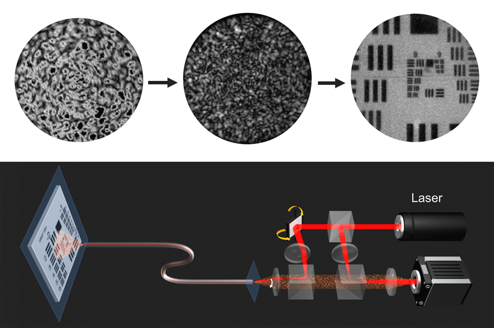
Now, to perform endoscopy, the authors have to exploit the transmission matrix twice (Fig. 1). First, on the way in, they illuminate the entrance of the fiber by various illumination angles, therefore illuminating an object placed just at the exit on the far side by well-determined, speckled illumination. Second, on the way out, the illuminated object reflects light back into the fiber, but the scrambled image that exits can be decoded, again thanks to the transmission matrix. The speckled nature of the illumination itself means that a single image is very noisy. However, changing the illumination multiple times (that is, by changing the angle at which the laser light enters the fiber) and summing the images, this speckled illumination averages out, and Choi et al. show that the result mimics an image illuminated by an incoherent source. The authors thus recover a very clean image, with an excellent resolution of 1.8 microns.
An extra advantage of the authors’ technique is that it is intrinsically equivalent to digital holography: the phase and the amplitude of the object are recovered. It is therefore possible to numerically reconstruct the object at different planes without physically scanning the fiber, hence obtaining information in all three dimensions. As a proof of principle, Choi et al. have performed preliminary images of tissue from rat intestine, demonstrating the resolution, field of view, and the multiple-plane imaging capability of their technique. While the measurement of a transmission matrix of a multimode fiber has been performed by other groups [6–8] and used for different applications such as tweezing, focusing, and fluorescence imaging [9], this work demonstrates, for the first time, the use of a multimode fiber as a real endoscope, where it is light reflected by the object that is imaged.
Deep imaging in complex media with optical contrast and micron resolution is of paramount interest, in particular for biomedical applications. Wave front shaping techniques, long known in radar and acoustics [10], have recently contributed to a new era in optics [11]. Today, the same techniques are helping improve endoscopy, making it significantly less bulky and invasive. Tomorrow, the same concepts might lead to new ways of performing high-resolution, noninvasive imaging of tissue at depths that are currently out of reach.
References
- Y. Choi, C. Yoon, M. Kim, T. D. Yang, C. Fang-Yen, R. R. Dasari, K. J. Lee, and W. Choi, “Scanner-Free and Wide-Field Endoscopic Imaging by Using a Single Multimode Optical Fiber,” Phys. Rev. Lett. 109, 203901 (2012)
- V. Ntziachristos, “Going Deeper Than Microscopy: The Optical Imaging Frontier in Biology,” Nat. Methods 7, 603 (2010)
- E. N. Leith and J. Upatneiks, “Holographic Imagery through Diffusing Media,” J. Opt. Soc. Am. 56, 523 (1966)
- I. M Vellekoop and A. P. Mosk, “Focusing Coherent Light through Opaque Strongly Scattering Media,” Opt. Lett. 32, 2309 (2007)
- S. Popoff et al., “Measuring the Transmission matrix in Optics: An Approach to the Study and Control of Light Propagation in Disordered Media,” Phys. Rev. Lett. 104, 100601 (2010)
- T. Čižmár and K. Dholakia, “Shaping the Light Transmission through a Multimode Optical Fibre: Complex Transformation Analysis and Applications in Biophotonics,” Opt. Express 19, 18871 (2011)
- S. Bianchi and R. Di Leonardo, “A Multi-Mode Fiber Probe for Holographic Micromanipulation and Microscopy,” Lab Chip 12, 635 (2012)
- I. N. Papadopoulos, S. Farahi, C. Moser, and D. Psaltis, “Focusing and Scanning Light through a Multimode Optical Fiber Using Digital Phase Conjugation,” Opt. Express 20, 10583 (2012)
- T. Čižmár and K. Dholakia, “Exploiting Multimode Waveguides for Pure Fibre-Based Imaging,” Nat. Comm. 3, 1027 (2012)
- M. Fink, ”Time Reversed Acoustics,” Phys. Today 50, No. 3, 34 (1997)
- A. P. Mosk, A. Lagendijk, G. Lerosey, and M. Fink, ”Controlling Waves in Space and Time for Imaging, and Focusing in Complex Media,” Nature Photon. 6, 283 (2012)


