Making a Molecular Movie with X Rays
The first step in the conversion of light into chemical or mechanical energy is the absorption of light by a molecule, resulting in a change of the molecule’s structure. The structure of the initial and final states of the molecule are usually known, but the transition between the two often happens so quickly that scientists have to rely on theoretical simulations to determine the dynamical structures. That is, one can see where an isolated molecule started and where it ended up, but one is left wondering how it got from one configuration to the other. This missing information is central to understanding how molecular reactions take place and to developing the tools to control them. Michael Minitti and colleagues [1] from SLAC National Laboratory, California, have now taken an important step towards imaging transient states by using femtosecond (fs) high-energy x-ray pulses to probe a reaction that takes place on a time scale on the order of 100fs.
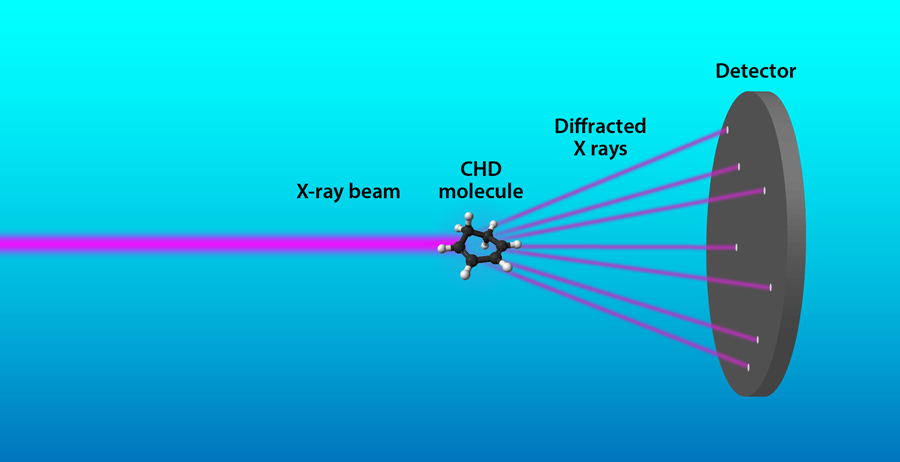
The field of femtochemistry [2] has been devoted to studying molecular reactions using spectroscopy with femtosecond laser pulses, which measures changes in the energy of the molecule as a function of time. It would be most useful to directly determine the structural dynamics of isolated molecules during a reaction. However, the challenge is to simultaneously achieve sub-angstrom spatial resolution to map the position of the atoms and sub- 100-fs time resolution to match the time scale of the dynamics. Diffraction experiments, either with electrons or x rays, can be used to measure the structure of molecules. As a wave scatters from different atoms in the molecule, these scattered waves will interfere on the detector. By analyzing this interference pattern, it is possible to obtain the distances between the atoms. Previously, electron pulses have been used to determine transient molecular structures in the gas phase because the scattering cross section is much higher for electrons than for x rays of comparable wavelength [3]. However, with the recent development of extremely bright hard-x-ray free-electron lasers (XFELs), it is now, in principle, possible to achieve atomic resolution with x-ray diffraction [4].
In their study, Minitti and colleagues used femtosecond hard-x-ray pulses to perform a time-resolved x-ray diffraction experiment on 1,3-cyclohexadiene (CHD) molecules (Fig. 1). The x-ray pulses were produced by the XFEL at the Linac Coherent Light Source at SLAC National Lab. The CHD molecule has a ring structure that opens after absorbing a photon of ultraviolet (UV) light on a time scale of 100fs. The research team investigated the photoreaction using a “pump-probe” experiment. The reaction was triggered with a femtosecond UV laser pulse (the pump), followed by an x-ray pulse (the probe) that is synchronized to the laser pulse. The x-ray pulse scatters from the sample, and the diffraction pattern is recorded on a detector. The authors repeated the experiment multiple times, using varying time delays between the laser and x-rays pulses to capture diffraction patterns at different moments of the reaction.
The XFEL pulses had a duration of only 30fs and an energy of 8.3 kilo-electron-volts that corresponds to a wavelength of 1.5 angstrom. The wavelength of the pulses is important because the maximum spatial resolution that can be achieved is approximately equal to the wavelength. In this experiment, the wavelength was too long to obtain the atomic structures directly from the diffraction patterns, but the team found an alternative method to elucidate the structure of the intermediate states in the reaction. The authors used numerical simulations to calculate the many possible paths that the molecules could take from the known initial state, given the energy of the UV photon that is absorbed.
To determine which trajectories actually take place in the experiment, Minitti and colleagues calculated diffraction patterns for all possible trajectories and compared them to the measured patterns. Using a mathematical tool known as nonlinear least-squares optimization, they found that only a few trajectories were needed to match the experimental results. Next, they deduced the atomic structures from the simulated trajectories that best matched the data. Armed with these structures, the researchers were then able to generate movies of the molecular reaction and to determine that the reaction took place in 80fs. The reaction time is consistent with previous spectroscopic measurements, but this experiment also provides information on how the shape of the intermediate states evolves in time.
These results open a new direction in the effort towards making molecular movies. While time-resolved electron-diffraction experiments have finer spatial resolution than that attained here, the present method has demonstrated higher temporal resolution, which is needed to be able to follow the motion of the atoms during a reaction. The next challenge for femtosecond hard-x-ray diffraction would be to improve the spatial resolution such that more details about the molecular structure can be learned, which can be reached by going to higher energy (shorter wavelength) and brighter x-ray pulses.
This research is published in Physical Review Letters.
References
- M. P. Minitti et al., “Imaging Molecular Motion: Femtosecond X-Ray Scattering of an Electrocyclic Chemical Reaction,” Phys. Rev. Lett. 114, 255501 (2015)
- http://www.nobelprize.org/nobel_prizes/chemistry/laureates/1999/
- H. Ihee, V. A. Lobastov, U. M. Gomez, B. M. goodson, R. Srinivasan, C. Y. Ruan, and A. H. Zewail, “Direct Imaging of Transient Molecular Structures with Ultrafast Diffraction,” Science 291, 458 (2001)
- P. Emma et al., “First Lasing and Operation of an Ångstrom-Wavelength Free-Electron Laser,” Nature Photon. 4, 641 (2010)


