Uncovering a New Layer

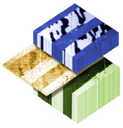
Computer hard drives and other advanced devices depend on layered stacks of magnetic and non-magnetic materials, but researchers don’t fully understand how the materials work at the microscopic level. In the 10 December print issue of PRL, a team describes a new technique for imaging the magnetic structures at the boundaries between these layers. Their data show that the boundaries are not as clean as previously assumed: a chemical reaction at the interface can cause an ultrathin layer of a new material to form. The work suggests that researchers need to look more closely at such interfaces in order to develop the next generation of devices.
Computer hard drives can cram 20 gigabytes onto a surface that previously held just one gigabyte, thanks to the high-tech use of layered magnetic materials. Condensed matter physicists are trying to further exploit these materials for devices such as transistors and light-emitting diodes that will be smaller, faster, and less power-hungry than today’s devices. Theoretical models of the materials assume there is no “leakage” of atoms between layers, but it’s difficult to analyze their chemical and magnetic properties at a microscopic scale to verify the assumption.
A team lead by Joachim Stöhr, of the Stanford Linear Accelerator Center in California, used a unique set of x-ray techniques to look at an important combination of materials that is not fully understood. A nickel oxide (NiO) layer on top of cobalt (Co) “freezes” the magnetic field direction in the Co, and although the effect has been used in devices for years, researchers don’t know exactly why it works.
The team used x-ray absorption spectroscopy (XAS) to find the spectrum of x-ray energies absorbed by the sample, which gives its chemical composition. They then imaged the sample with x-ray photoemission electron microscopy (XPEEM), a recently developed technique that gives a microscopic map of the magnetic structure. In XPEEM, x rays hit the sample and eject electrons, which are imaged by an electron microscope. Circularly polarized x rays preferentially eject electrons from atoms magnetically aligned with the polarization axis. The team also used linearly polarized x rays to learn about the magnetic structure. They found that an ultrathin layer of NiCoO had grown between the NiO and Co, produced by a chemical reaction between the layers. The reaction leaves some pure nickel atoms behind, and they provide a strong enough magnetic field to freeze the sandwich’s total field.
Hendrik Ohldag, a member of the Stanford team, hopes the results will encourage others to take a closer look at similar interfaces in other materials. Even though chemical reactions will not always occur, he suspects other interface effects may be found. Yves Idzerda of Montana State University in Bozeman agrees that these x-ray techniques are likely to reveal additional information regarding the nature of magnetic interfaces. “This will lead to improved control, reliability, and tunability of magnetic device structures,” he says.
–Tim Palucka
Tim Palucka is a freelance science writer in Pittsburgh.


