Quick Pictures with Electrons
Electron microscopes use narrow beams of energetic electrons to image objects too small to be seen with light, such as atoms in a semiconductor or cellular organelles. Researchers have adapted the microscopes to capture fast changes in materials: they take a series of quick images, each with a short, intense pulse of electrons—the equivalent of taking photographs with a brief, bright flash. But these microscopes often have to trade spatial resolution for speed: a shorter pulse has to carry more electrons to produce a sharp image, and repulsion between the charges causes the beam to spread.
Now, researchers at the University of California, Los Angeles, propose an electron microscope design that—at least in theory—is capable of capturing, in a single shot, images of -nanometer-sized objects within picoseconds ( seconds)—about times faster than the highest-speed microscopes in operation today. As reported in Physical Review Applied, the proposed microscope would allow researchers to study how shock waves or temperature gradients affect a material’s structure.
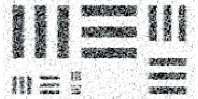
The key component in Renkai Li and Pietro Musumeci’s proposal is a relativistic electron source, called a radio-frequency photoinjector, in which electrons are stripped by a laser from a metal cathode and quickly accelerated to relativistic energies. These sources, which are similar to those used to seed some x-ray free-electron lasers, are capable of emitting high peak currents in tight bunches. Moreover, relativistic electrons experience a lower charge density in their rest frame (because of time dilation). Li and Musumeci’s simulations show that an electron microscope with a photoinjector source and special quadrupole magnet lenses is able to image a test pattern of nanometer-sized bars within picoseconds. – Jessica Thomas


