Magnetic Cells
Laboratory-grown cell aggregates (“spheroids”) are used as model systems to simulate how biological tissues grow and develop, and to give insight into their mechanical properties. Now, Claire Wilhelm at Paris Diderot University, France, and colleagues have developed a new method, based on embedded magnetic nanoparticles, for engineering and characterizing spheroids. Unlike other techniques, their method allows large millimeter-sized spheroids to be formed, real-time imaging, and the characterization of multiple mechanical properties, including surface tension, viscoelasticity, and yield stress.
To provide their cells with magnetic properties, the group added magnetic nanoparticles to the cell culture medium. The cells absorbed these nanoparticles with no alteration to their functions. Filling a mold with these cells and turning on a magnetic field, the authors were able to compact the cells, forming a spheroid with uniformly distributed nanoparticles. The technique yielded spheroid sizes up to few millimeters, much larger than those attainable by previous methods.
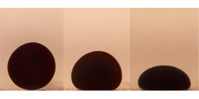
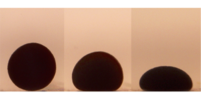
To probe the spheroids’ mechanical properties, the authors placed them on a flat surface and flattened them by applying a permanent magnetic field. The experiments revealed that small and large spheroids responded differently to flattening: Small spheroids behaved like liquid drops, while larger spheroids behaved like elastic solids at short times and like liquid drops at longer times. The setup could be enhanced to carry out time-dependent measurements by using an electromagnet: An oscillating magnetic flattening force would allow the viscoelastic response of the spheroids at different frequencies to be probed. This technique could also be used to study how processes like cell division or cell death change the mechanical properties of a tissue, and to characterize other soft systems, such as hydrogels or emulations.
This research is published in Physical Review Letters.
–Katherine Wright


