A TEM That Images Quantum Currents
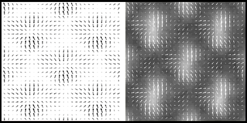
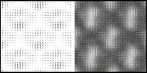
Transmission electron microscopes (TEMs) allow scientists to see individual atoms or structures inside a biological cell. Axel Lubk, at the University of Dresden in Germany, and colleagues have added to these capabilities by reconfiguring a commercial TEM so it can be used to map crystalline strain and electric or magnetic fields within a material. The crux of their approach is to image the quantum-mechanical electrical currents associated with the scattering of the TEM electron beam in the material.
TEM imaging involves passing a narrow electron beam through a sample and detecting a modulation in its intensity. The effect depends on the charge density the beam encounters in the sample, allowing (with certain microscope configurations) a map of a crystal’s atoms to be obtained. But the interaction between the beam and the sample also generates quantum-mechanical currents. And compared to a map of the beam’s intensity, a map of these currents can be more sensitive to crystalline strain or a jump in magnetization near a domain wall.
So far, detecting these currents has required a coherent electron beam, which complicates the setup. Lubk and his colleagues get around this constraint. Using an incoherent beam, they compare a series of unfocused images, acquired at increasing distances from the sample. Because of the currents associated with the scattering electron beam, each image is slightly different. By comparing three such images, the team could reconstruct the currents in a thin film of strontium-titanate with atomic resolution. A time series of these current maps could be used, for example, to track nanoscale regions of strain that occur in a thin vibrating membrane.
This research is published in Physical Review Letters.
–Jessica Thomas


