3D Images 10 Times Faster
In x-ray phase contrast imaging (XPCI), pictures are created by tracking changes in both the x rays’ phase and absorption after they have passed through the object of interest. Now researchers report a new image reconstruction algorithm that halves the number of x-ray shots required to produce the image. Implementing the algorithm also eliminates the need to reposition optical elements between exposures, so the total acquisition time can be cut to a tenth of the usual. Faster acquisition may benefit biological studies that often have a limited time window.
XPCI can provide more detailed images than traditional x-ray techniques because it tracks both the absorption and the phase changes induced as x rays pass through an object. In one version of XPCI, an x-ray beam illuminates an object sandwiched between two misaligned masks—before and after the object—each consisting of an array of vertical slits. A detector records the intensity of x rays that make it through the sandwich. The slits make the technique sensitive to sideways deviations in the x rays’ routes through the object (refraction) due to density variations. And since refraction is a result of the waves’ phase variation, the mask allows phase effects to be detected.
To create a 2D snapshot, the object is imaged twice, with the slits in two slightly different positions. That’s because two shots are needed to disentangle the effects of refraction and absorption. And for 3D images, this process has to be repeated many times as the object is rotated. The procedure is relatively simple, but the constant moving of the masks is time consuming.
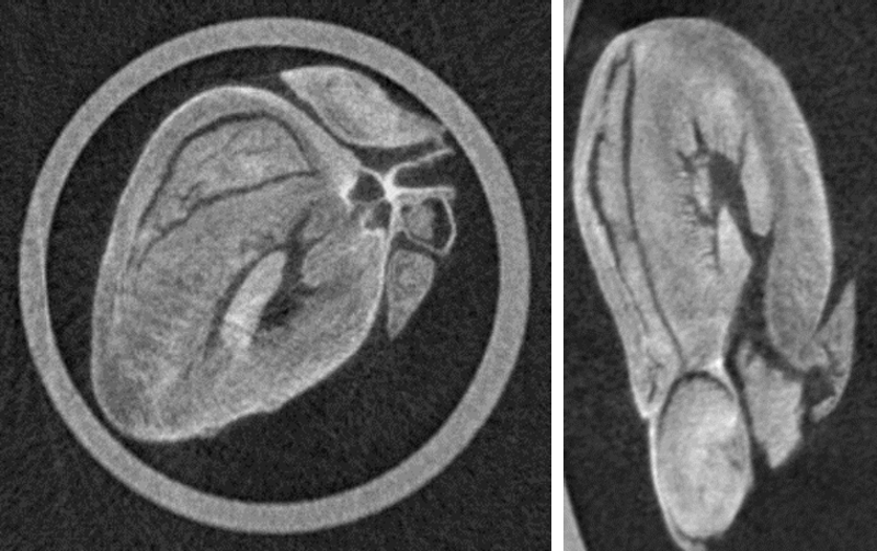
Paul Diemoz from University College London and colleagues realized that if they assumed that the ratio of the attenuation coefficient (which represents absorption) to the index of refraction remains constant—a good approximation in many biological samples, for example—then they could get away with a single exposure for each 2D slice. The team tested their method by imaging an array of plastic tubes, a beetle, and a freeze-dried rat’s heart, showing that the technique works for both artificial and biological objects. With their best x-ray detector, the researchers took about 3.5 minutes to collect the 3D heart data, nearly 10 times faster than the previous best of 25 minutes. Such fast acquisition could be useful for imaging patients, who can only lie still for a limited time.
This research is published in Physical Review Applied.
–Katherine Wright
Katherine Wright is the Deputy Editor of Physics Magazine.