X-Ray Probe Targets Interfaces
Interfaces are what separate one material from another. We sense the world through interfaces, whether touching the surface of a table or seeing the light reflecting off the edge of a glass. Many other interfaces are less visible but still have a place in our lives. Modern solar cells consist of thin layers where interfaces play an important role for charge separation. Catalytic reactions for chemical energy transformations occur at solid-gas or solid-liquid interfaces. It is extremely challenging to probe these interfaces, as they are often buried under layers or in contact with a liquid or high-pressure gas where the number of interface atoms is extremely small in comparison to the surrounding material [1]. Researchers, therefore, search for techniques that are only sensitive to the two-dimensional interface. Royce Lam from the University of California, Berkeley, and colleagues have developed an interfacial probe utilizing soft-x-ray pulses from a free electron laser [2]. By aiming the pulses at a graphite sample, the scientists have shown that they can detect a nonlinear spectrographic signal that arises from the graphene layers near the surface of the graphite. This demonstration opens up a new field in interface studies, offering the possibility to track surface chemistry reactions with the femtosecond resolution provided by the very short x-ray pulses from free electron lasers.
The new interfacial probe relies on a process where two photons of the same energy are absorbed by an atom to generate a photon with double the original energy [3]. This so-called second harmonic generation (SHG) can only occur in regions of a material that are asymmetric—specifically, regions that lack inversion symmetry. This asymmetry is necessary to counter interference effects that would cancel out the SHG emission. An interface is by nature asymmetric, which is why researchers have for many years used SHG observations to investigate interfaces based on optical laser techniques [4]. Having a similar capability in x rays would allow measurements of the electronic structure using core level spectroscopy, where the inner shell provides atom-specific sensitivity [5]. However, x-ray-based SHG studies have lagged behind those in the optical regime due to a lack of sources for coherent, high-intensity x rays, which are needed to generate multiple absorptions. That has begun to change with the development of free electron lasers (FELs). Previous FEL experiments in the hard-x-ray regime (between 10 keV and 100 keV) have observed SHG, but the effect has so far not been localized to the interface [6, 7].
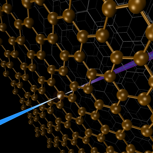
The work by Lam et al. is one of the first to investigate SHG with soft x rays (between 100 eV and 1 keV). The research was performed at the Free Electron laser Radiation for Multidisciplinary Investigations (FERMI) facility in Trieste, Italy [8], which has recently increased its beam energy to allow measurements at the inner shell carbon 1s level, denoted by the K edge (284 eV). The absorption at the K edge is a resonant process corresponding to the excitation of electrons from an atom’s innermost orbital to one of its unoccupied outer orbitals. In the experiments, the team focused short soft-x-ray pulses of 25 femtoseconds on a graphite sample (see Fig. 1). The carbon atoms absorbed some of these x-ray photons and re-emitted photons of characteristic energy as they relaxed back to their ground state. The researchers recorded this emission, as well as the transmitted light, with an x-ray spectrometer. They measured the fundamental intensity, which is the output corresponding to single-photon absorption, and used that to normalize the SHG signal, which is the intensity at twice the fundamental energy.
In a series of laser shots, the researchers varied both the incoming laser-pulse energy and the graphite thickness. For each pulse, they recorded the SHG intensity at three different excitation energies around resonances in the graphite x-ray absorption spectrum. Specifically, one excitation energy was below the carbon K edge, another was around the excitation into the first unoccupied orbital and the last was in the ionization continuum. The SHG intensity exhibited a nonlinear dependence with pulse energy, indicating a two-photon process. The SHG signal was only detected for electronic processes involving x-ray absorption—that is, above the K edge. Since this signal was quite strong, SHG detection should be possible with lower pulse intensities, which will be important for more fragile samples that otherwise could be destroyed in the intense x-ray laser beam.
The most significant finding for future utilization is the observation that the SHG signal is strongly enhanced upon resonant excitation into the unoccupied states in graphite. This means that interface-selective x-ray absorption spectra could be measured in SHG mode. X-ray absorption spectroscopy is already a well-established technique for studying chemical bonding and structure of species on surfaces [9]. By targeting the SHG signal in an x-ray absorption spectrum, researchers could identify the presence of particular reaction products on the solid-liquid or solid-gas interface of a catalyst during operating conditions. Combining this soft-x-ray probe with an optical laser to pump the system would allow researchers to follow surface chemical reactions in real time with femtosecond resolution [10]. This will be essential in the development of new catalysts for the chemical industry, but it may also help with efforts to improve fuel cells or to create artificial photosynthesis systems.
This research is published in Physical Review Letters.
References
- G. A. Somorjai, Introduction to Surface Chemistry and Catalysis (John Wiley & Sons, Inc., New York, 2010)[Amazon][WorldCat].
- R. K. Lam et al., “Soft X-Ray Second Harmonic Generation as an Interfacial Probe,” Phys. Rev. Lett. 120, 023901 (2018).
- Y. R. Shen, “Surface Properties Probed by Second-Harmonic and Sum-Frequency Generation,” Nature 337, 519 (1989).
- P. A. Franken, A. E. Hill, C. W. Peters, and G. Weinreich, “Generation of Optical Harmonics,” Phys. Rev. Lett. 7, 118 (1961).
- A. Nilsson, “Applications of Core Level Spectroscopy to Adsorbates,” J. Electron Spectrosc. Relat. Phenom. 126, 3 (2002).
- T. E. Glover et al., “X-Ray and Optical Wave Mixing,” Nature 488, 603 (2012).
- S. Shwartz et al., “X-Ray Second Harmonic Generation,” Phys. Rev. Lett. 112, 163901 (2014).
- E. Allaria et al., “Highly Coherent and Stable Pulses from the Fermi Seeded Free-Electron Laser in the Extreme Ultraviolet,” Nat. Photon. 6, 699 (2012).
- J. Stöhr, NEXAFS Spectroscopy, edited by G. Ertl, R. Gomer, and D. L. Mills, Springer Series in Surface Science (Springer-Verlag, Berlin-Heidelberg, 1992)[Amazon][WorldCat].
- H. Öström et al., “Probing the Transition State Region in Catalytic CO Oxidation on Ru,” Science 347, 978 (2015).


