Q&A: Using Quantum Tricks to Scan the Brain
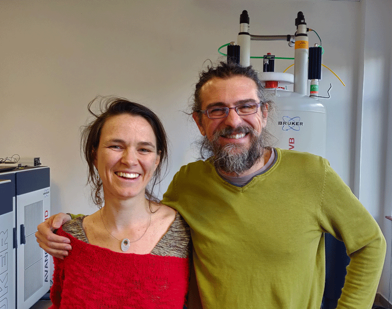
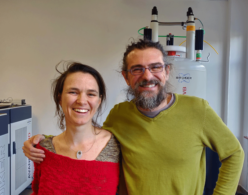
Twenty years ago, researchers figured out how to detect the early stages of a stroke using a technique called diffusion magnetic resonance imaging (dMRI). Today, quantum information experts Analia Zwick and Gonzalo Álvarez from Argentina’s Bariloche Atomic Center (CAB) are using their skills to unlock more information from each pixel of a dMRI scan. Although their research is still in the experimental stage, they hope it might lead to earlier diagnoses of diseases that affect the brain, like Alzheimers.
The two physicists, who are married to each other, began their careers finding ways to protect atomic spins and other delicate quantum systems from their surroundings. The dMRI technique that they are developing turns that idea around and uses the spins of water molecules as quantum sensors of the body.
Zwick and Álvarez recently returned to Argentina to set up a lab at CAB, a research campus in the picturesque lake region of Patagonia. They proudly own South America’s strongest magnet for pre-clinical medical imaging, and they are part of a national effort to bring physics and medicine together. In an interview with Physics, they explained how they are pushing the limits of MRI sensitivity and why planting roots in their home country was always their goal.
–Jessica Thomas
You are using quantum tricks to enhance dMRI. Before getting to that, could you explain how conventional dMRI works?
Gonzalo: The dMRI technique is sensitive to the movement of water molecules in the body. The brightest parts of the image correspond to molecules that move the least during a scan. The molecules move by diffusion, and this movement depends on the type of tissue the molecules are in or whether they are restricted, say by cell walls.
How do you see these motions?
Analia: In conventional dMRI, we subject the sample to two magnetic field pulses separated in time. The first pulse applies a field gradient and causes the spins of the sample’s water molecules to precess at a rate that depends on where they sit in the sample. The second pulse reverses the gradient and the precessions. If the molecules haven’t moved, they rewind to where they started.
Each precessing spin acts like a “runner” racing around a track. The first field pulse makes the runner run one way along its track; the second pulse asks it to come back. But the runner will only return to its starting position if it stays on its original track, that is, if the molecule hasn’t moved in the sample. The signal we measure depends on the number of spins that return to their starting position. From that signal, we can infer how much the molecules move.
Why do you want to enhance the technique?
Gonzalo: The problem with conventional dMRI is that it doesn’t tell you why the molecules move fast or slowly. To get this sensitivity, our group and a few others use so-called “quantum control” techniques that were originally designed to protect quantum information.
How do you fold in this “control”?
Analia: We play with the instructions that we give to the spins. So, for example, we change when they should go forward then backward, or how often they should repeat these steps. The instructions are designed so that we can extract as much information as possible about why certain running spins don’t come back to where they started.
What would this enhanced technique allow you to study?
Gonzalo: We can already measure the diameters of cells and the distribution of these diameters. The sizes of brain cells, for example, change depending on their function, or on their health. But the changes are so slight, you can usually only see them by taking a slice from the brain and putting it under a microscope. We believe that our enhanced techniques will allow cell changes to be measured inside the patient and noninvasively.
Analia: Quantitative information from dMRI images might help in the early detection of some illnesses. For example, the brain’s axons are covered in a layer of myelin—like electrical cable is coated in plastic. In Alzheimer’s disease, the myelin gets damaged. Seeing signs of this damage could potentially allow for earlier diagnosis of the disease.
Both of you come from a fundamental physics background. What inspired you to switch gears and focus on medical applications?
Gonzalo: When I was a Ph.D. student, I thought that physics was about answering the question “Why is this happening?” But then I did research at the Weizmann Institute with Lucio Frydman, a leading NMR scientist. He always tries to see how he can make something useful. I realized that scientists don't only need to answer questions that start with “Why” but also those that start with “What for.”
Analia: People like to say we’re now in the second quantum revolution, where we try to control and manipulate quantum information for useful applications, such as nano- or molecular-scale sensors. We started to do exactly that during our postdocs abroad, and then we had the opportunity to return to Argentina and set up a lab in a new medical physics department.
Gonzalo: This opportunity was la frutilla del postre—an expression we have in Argentina that means “the strawberry on the cake.”
How have you found the return?
Gonzalo: We always intended to come back. We wanted to go abroad, learn some things, and then return to Argentina and help develop the country.
There is a lot of work to do—we’re not only doing our research, but also helping to develop institutions and the way science is done in Argentina. This combination is challenging, because you have to do many things at the same time and still be competitive in research. But it’s very motivating to help develop your country and to help create a new path for the future generation.
Jessica Thomas is the Editor of Physics.
Know a physicist with a knack for explaining their research to others? Write to physics@aps.org. All interviews are edited for brevity and clarity.


