Mapping the Textures of Thicker Magnets
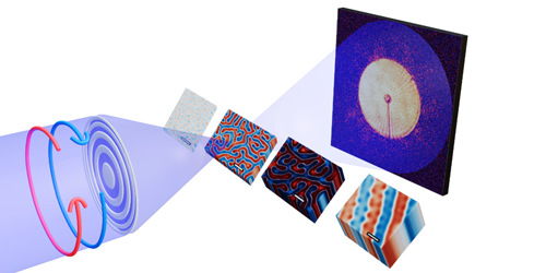
Understanding magnets’ nanoscale structures is critical to developing magnetic materials for applications in clean energy, sensing, computing devices, and many other technologies. While x-ray and electron microscopy can create high-resolution images of magnetic thin films, imaging thicker samples is often impossible. Jeffrey Neethirajan of the Max Planck Institute for Chemical Physics of Solids in Germany and his colleagues now overcome this limitation with an x-ray imaging technique for micrometer-thick magnets [1].
Using a soft-x-ray beam line at the Diamond Light Source, the UK’s national synchrotron facility, the researchers scanned a circularly polarized x-ray beam across a magnetic sample and then repeated the process with the polarization reversed. Circularly polarized x-rays interact differently with magnetic structures depending on their polarization direction—an effect called x-ray magnetic circular dichroism (XMCD). By comparing diffraction patterns obtained using each polarization direction, magnetic structures inside a sample can be mapped.
The conventional version of this technique harnesses this polarization-dependent effect by measuring differences in the way the two beams are absorbed. But this absorption signal only shows up across a narrow range of x-ray energies at which the sample is highly absorbing. This means that only thin samples, which these x rays can penetrate, can be imaged. Instead, Neethirajan and his colleagues extracted the XMCD phase information. This phase signal exists for a wide range of energies, making it possible to image thicker samples using x-ray wavelengths that are less strongly absorbed.
The researchers demonstrated the technique for specially fabricated samples up to 1.7 µm thick—an order of magnitude thicker than absorption XMCD can manage. They also imaged chiral magnets and naturally magnetic minerals, whose magnetic textures were previously inaccessible. They say that the capability opens the door to characterizing magnetic materials with applications in energy harvesting and spintronics.
–Rachel Berkowitz
Rachel Berkowitz is a Corresponding Editor for Physics Magazine based in Vancouver, Canada.
References
- J. N. Neethirajan et al., “Soft x-ray phase nanomicroscopy of micrometer-thick magnets,” Phys. Rev. X 14, 031028 (2024).