Watching a Virus Begin Its Attack
For a virus to infect a cell, it must first attach to the cell’s surface. A study in Physical Review Letters describes a new technique for measuring the strength of the virus-cell bond in a more direct way than conventional methods. The experiment uses a microscope to detect the continuous binding and unbinding of individual viruses. The technique may help researchers develop treatments for viral infections, such as anti-viral drugs that interfere with the attachment process.
Viruses consist of genetic material (DNA or RNA) contained in a protein shell. To infect a cell, a virus must first attach its protein coat to the cell membrane. Some antiviral drugs block the attachment process for specific viruses, but for many viruses, researchers don’t have enough information about the details of the process.
For example, measurements of the energy barrier a virus faces on binding and the strength of the virus-cell-membrane bond can be challenging. They usually involve measuring the time it takes for many virus particles to bind or unbind from cell membranes. But the measured rates are often dominated by the speed at which the particles diffuse through water, rather than by the details of the binding process, so these experiments can be hard to interpret.
Fredrik Höök of the Chalmers Institute of Technology in Sweden and his colleagues devised a technique where the binding and unbinding are in equilibrium—there is no net change over time in the number of bound viruses—and the diffusion rate has no effect. They demonstrated it with human norovirus, a gut infection that causes diarrhea, vomiting, and stomach pain and that wreaks havoc on cruise ships and in other enclosed environments.
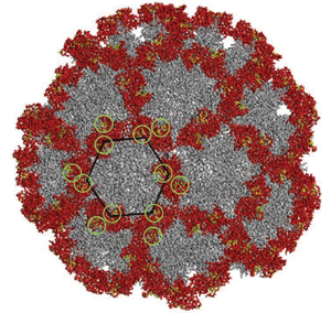
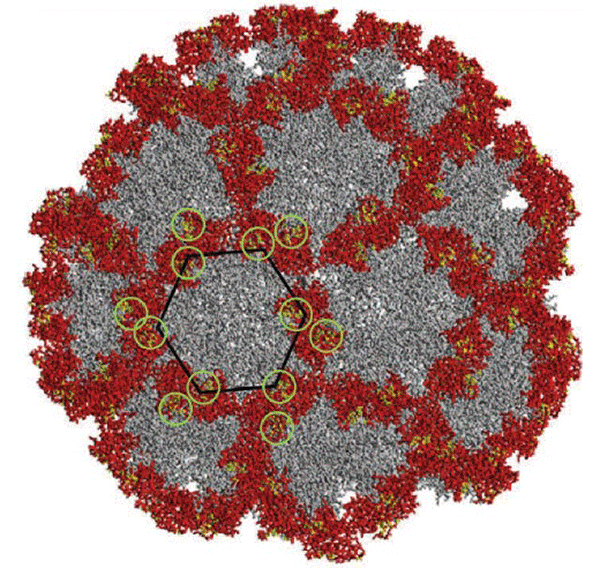
Since norovirus cannot be cultured in the lab, the team used only its protein shell in their experiments. They immobilized these “virus-like particles” (VLPs) on the bottom of a Petri dish and introduced lipid vesicles—spheres of cell membrane material—into the fluid above. The vesicle membranes were made primarily of conventional lipids but also contained fluorescent dye and a dash of molecules called glycosphingolipids, which are known to be involved in virus attachment to cells. The researchers used a total internal reflection fluorescence microscope, which detected fluorescence within only about of the bottom of the dish. Vesicles appeared as bright spots when they drifted close to the VLPs, but a spot only remained for significant time if the vesicle became attached to a VLP.
The team took several sets of time-lapse photos, each with different time intervals, and calculated the rates of binding and detachment of the vesicles, from which they derived the energies for binding and unbinding. They used vesicles with two different types of glycosphingolipids and were able to distinguish between them based on their detachment rates, suggesting that the technique could help researchers follow the virus’s rapid evolution, the team says. The researchers also noticed that there was not a single detachment rate, but a wide range of rates, which they attributed to differences in the number of virus-membrane bonds from one VLP to the next—possibly a common situation in real virus attachment.
“This is a gateway for new drug development,” says Christian Eggeling of the Max Planck Institute for Biophysical Chemistry in Göttingen, Germany. “It is important to find rate constants for every single step [of the binding process].” Höök says his team hopes their method will be used to study the evolution of norovirus by looking back at old preserved blood samples taken from patients over a period of decades. They also hope it will lead to the development of compounds that could prevent norovirus from attaching to cells.
–Catherine Lussenhop
Catherine Lussenhop is a freelance writer in Minneapolis.
References
- Manish M. Patel et al., Emerg. Infect. Dis. 14, 1224 (2008)


