Photoreceptors Measure Photon Statistics
For many years, people have tried to understand the structure of vision and its role in the perception of the world by living organisms. Suffice it to say that, thanks to their visual organs, people receive most of their information about the world by optical means. Eyes of living organisms represent advanced light harvesting systems, developed through hundreds of millions of years of evolution. Some of their features are comparable or even superior to existing man-made photodetection devices. For example, rod photoreceptor cells of the retina, which are responsible for night vision, are small photodetectors, containing a photosensitive element (rhodopsin pigment) along with a “built-in” chemical power supply (adenosine triphosphate produced by mitochondria). Remarkably, these photodetectors have very high sensitivity, down to the single-photon level.
As a result of this high sensitivity, statistical features of the photon fluxes (or, in other words, probability distribution of numbers of photons in each detection event) generated by different light sources might be monitored by these natural biological photodetectors. A crucial step towards a deep understanding of photoreceptor functioning would be a careful characterization of their response to well-controlled classical light sources. Such a study forms the focus of the work reported in Physical Review Letters by Nigel Sim and colleagues at the Data Storage Institute in Singapore [1], and it can be considered a pioneering research effort performed at the interface between physics and biology.
Many worthy experiments related to the sensitivity of the isolated rods have been performed in the past. However, either the statistics of rod response was not of interest to those researchers, or the noise properties of individual rods were difficult to assess since several hundred rods were simultaneously illuminated, so their collective response undergoes several intermediate stages of visual processing.
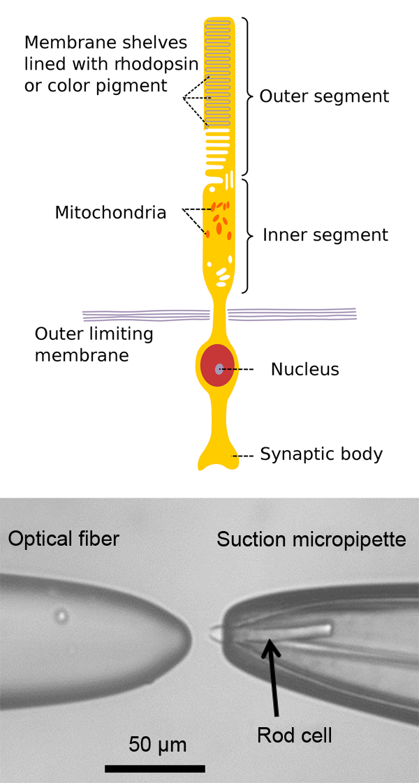
The objects under study are vertebrate retinal photoreceptor rod cells, which are densely packed in the retina in a layer covering the inner surface of an eyeball. Individual rods consist of two distinctive functional regions: an extended rodlike outer segment, which is filled with rhodopsin photopigment molecules, and a shorter, rounded, inner segment containing various components of cell machinery (Fig. 1, top). In the dark, the electrochemical gradient across the rod cell membrane causes a continuous ion flux in and out of the cell through aqueous pores in the membrane. Photons, impinging on the outer segment, cause reconfiguration of a rhodopsin molecule, which initiates a series of intracellular reactions. A fraction of the ion channels close, resulting in a sort of polarization of the cell and excitation of a signal to the optic nerve. Illumination with a relatively bright flash closes all light sensitive channels in the outer segment and bleaches the rod. A recovery process regulates reopening of the ion channels to the resting state.
The current experiments were performed with a so-called suction-electrode technique [2]. In this case, the outer segment of the rod is drawn into a tight-fitting glass microelectrode filled with a solution that reproduces physiological conditions. The ion current through the membrane is redirected to a low-noise amplifier and then is properly detected. It is important to stress that the tested rod works in a way similar to the in vivo system. Of course, this technique and the experiments described in the work [1] do not answer the question about the role of the brain in processing information collected by the biological receptor. However, Sim et al. obtained a clear answer to the question of the fundamental possibility of distinguishing between different types of light statistics by such detectors.
The technique described above allows the authors to study the influence of controllable photon fluctuations on the response of an isolated rod in the whole dynamic range of its response. To create the stimulus, they used two types of light sources with different statistics (a coherent laser beam and a “pseudothermal” source obtained by sending the same laser beam through a rotating ground-glass disk). Indeed, the coherent statistics serve, in some sense, as a reference because the average number of photons coincides with its variance, while the photon distribution with thermal statistics tends to bunching, which gives rise to “excess” correlations. The experimental results revealed a crucial influence of the bleaching of the rod on the fluctuations of its response.
In the experiments, the light beam coming from the selected source was sent into a standard Hanbury Brown and Twiss interferometer (this interferometer consists of a beamsplitter that equally distributes the initial beam between two output arms) coupled with a pair of fibers. One of the fibers is connected to a regular avalanche photodiode detector and the other stimulates the rod. With this setup, Sim et al. were able to obtain intensity correlations for the different light sources from the joint measurement of the rod and a reference detector. The retinas were taken from the dark-adapted eyes of adult male frogs (Xenopus laevis) and sliced with a blade to release single rods. The rods were loaded into a solution-filled chamber in a microscope, and a glass micropipette was used for conventional suction-electrode recordings. The outer segments of intact rods were captured in the pipette by gentle air suction (Fig. 1, bottom). The photocurrent pulses were amplified, digitized, and analyzed by computer.
Sim et al. analyzed their data, and under some reasonable assumptions, found good agreement between theory and experiment. In particular, each photon isomerizes only a single rhodopsin molecule and in the absence of saturation, subsequent electrophysiological responses to individual isomerizations are additive and have a standard Gaussian shape.
The research performed by Sim et al. [1] reveals how the normalized average photocurrent amplitudes and the second-order intensity correlation function depend on the average numbers of photons for coherent and pseudothermal sources. These dependences demonstrate that saturation of the photocurrent amplitude for the coherent source is significantly steeper than for the pseudothermal one. This can be explained by the influence of the excess correlations inherent in the thermal statistics (in comparison with the coherent one) on the response of the individual rods. Thus correlations of the pseudothermal source are gradually neglected due to the saturation of the rod response. Indeed, for the pseudothermal source, unlike the coherent source, the probability of occurrence of a relatively low number of photons remains non-negligible, even for a large value of the average. This results in the widening of the amplitude probability distribution and prevents sharp saturation of the response. Moreover, at a relatively low number of impinging photons, the statistics of the response is not affected by the saturation for both cases. However, when the rod is stimulated by bright flashes, the probability of the saturation amplitude becomes dominant and causes damping of the amplitude fluctuations. For the rods studied in the experiments, the typical number of photons per light pulse, which causes the saturation, was about one thousand.
With their results, the authors have shown that these biological receptors act, in some sense, like commercially available photodetectors used for quantum optics and quantum communication because they are able to distinguish between light sources with different statistical properties. In spite of the fact that only classical light sources have been tested, these experiments clearly demonstrate the ability of receptors to respond to the photon statistics, and thus testing sources of quantum states of light would be the next logical step. Another important direction would be clarifying the role of the brain in processing the data coming from such receptors.
The work of Sim et al. [1] serves as a starting point for further investigation of biological sensors illuminated by nonclassical light fields. The results of such research could be motivation for building a whole family of advanced devices based on rod photoreceptors as well as fostering wider use of physical methods (including those used in quantum information/communication) in the study of biological structures.
References
- N. Sim, M. F. Cheng, D. Bessarab, C. M. Jones, and L. A. Krivitsky, ”Measurement of Photon Statistics with Live Photoreceptor Cells,” Phys. Rev. Lett. 109, 113601 (2012)
- D. A. Baylor, T. D. Lamb, and K.-W. Yau, “The Membrane Current of Single Rod Outer Segments,” J. Physiol. 288, 589 (1979)