Tiny Lasers Could Create High-Resolution Images
The resolution of images made with a standard optical microscope is usually limited to about a wavelength of light. But what if the light source is smaller than that? Researchers have now demonstrated a technique that could produce images from the light emitted by microscopic particles acting as tiny lasers. By embedding a large number of these emitters in a sample such as a biological cell, the researchers say it should be possible to make an image with a resolution comparable to the size of the particles, which are smaller than a wavelength. The method could be particularly useful for deep imaging of biological tissues.
The phenomenon of diffraction means that light coming from two sources can’t generally be distinguished if they are closer than about half the light’s wavelength. Obtaining optical resolution smaller than the wavelength of the light is thus said to beat the diffraction limit. For visible light, this means a few hundred nanometers.
Various imaging techniques for beating the diffraction limit already exist. One of them, laser confocal microscopy, manipulates the light waves to illuminate, say, a sample of biological tissue, one subwavelength spot at a time, where it induces fluorescent emission from a dye that was previously added to the tissue. Scanning the cell in this way yields an image with subwavelength resolution, but unwanted emission from cell components other than the dye reduces the sensitivity of the method and makes it ineffective for thick samples.
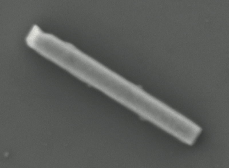
The new approach reported by Seok Hyun Yun and co-workers at the Massachusetts General Hospital and Harvard Medical School in Cambridge, Massachusetts, overcomes these limitations. The researchers made tiny lasers in the form of short rods (nanowires) of a crystalline compound called lead iodide perovskite, just 3–7 micrometers ( m) long and 300–500 nanometers (nm) across. This material acts as a semiconductor laser (see Focus Landmark: Invention of the CD-Player Laser) and can be “pumped” with green light to stimulate laser emission at a wavelength of 775 nm.
Yun and colleagues placed nanowires in a dilute suspension on a glass slide. Scanning with a probe beam 2.5 m wide, they saw emission turn on and off abruptly as the probe crossed individual nanowires. The emitted light was of a single wavelength, and it switched on suddenly as the intensity of the probe beam crossing a nanowire rose above a certain threshold for pumping. The researchers concluded that they were seeing genuine laser emission from the nanowires.
In principle, the position of the probe beam can be controlled with a precision of tens of nanometers, the researchers say, allowing the edges of the nanowire to be located with the same precision—well below the diffraction limit. It should be possible to resolve lasing particles just 50 nm wide, the researchers say.
To use the technique for imaging, researchers would “stain” specific structures within a sample with lasing nanowires (or even smaller nanoparticles such as quantum dots), so that these could be identified in the images. Attached to organelles or protein assemblies inside cells, say, such particles should allow these structures to be tracked with 10-nm resolution. “I envision laser particles that are injectable, implantable, or even swallowable in experimental animals, for biomedical investigations,” Yun says.
The new technique should be especially suitable for deep imaging within thick samples, such as tissues, because the intense, single-wavelength laser light can easily be distinguished from diffuse background fluorescence. “We aim to demonstrate the full potential of the technology for deep imaging,” says Yun.
“This is a clever concept,” says Malte Gather of the University of St. Andrews in the UK. But he adds that so far it is a preliminary proof of principle—the resolution demonstrated was no better than that of conventional, state-of-the-art optical microscopy. He agrees with Yun and his colleagues that smaller laser particles will be needed for high-resolution imaging. To get a detailed image of a complex sample, says Gather, “one would have to stain it with billions of lasers”—which would also require that they be much smaller.
This research is published in Physical Review Letters.
–Philip Ball
Philip Ball is a freelance science writer in London. His latest book is How Life Works (Picador, 2024).