Taking Pictures with Single Ions
Ions and electrons are often used in microscopy because they can provide much higher resolution than visible light. However, at high flux rates, these charged particles can adversely affect or damage a sample. Georg Jacob, from the University of Mainz in Germany, and colleagues have now developed an ion microscope that works with just one ion per image pixel. As a demonstration, the researchers used the device to take images of perforated objects with nanometer resolution.
In normal ion transmission microscopy, a source emits ions that are accelerated towards a target and detected if they are transmitted. Both the source and the detectors are noisy, so researchers typically boost ion flux to improve the signal-to-noise ratio of the imaging. The problem is that many targets, such as biological samples, cannot withstand high ion exposure.
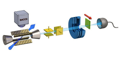
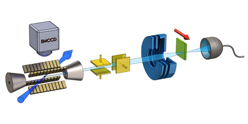
The ion microscope designed by Jacob and co-workers achieves a high signal-to-noise ratio while minimizing the ion flux rate. The method begins with capturing calcium ions in a one-dimensional electromagnetic trap. Applied electric fields extract single ions from one end of the trap and accelerate them towards a nanometer-sized focus region, through which a target object is moved incrementally. At each incremental step or “pixel,” a single ion emitted from the trap is either blocked by the object or passes through to a detector. Because the ion emission time is determined precisely, the researchers can turn off the detector, except when the ion is expected to arrive, thus reducing by a millionfold the noise from “dark counts” in the detector. With less than 600 ions, the team was able to determine the position of a 1- m circular hole in a diamond sample with an accuracy of 2.7 nm.
This research is published in Physical Review Letters.
–Michael Schirber
Michael Schirber is a Corresponding Editor for Physics based in Lyon, France.


