Landmarks: NMR–Grandmother of MRI
APS has put the entire Physical Review archive online, back to 1893. Focus Landmarks feature important papers from the archive.
Magnetic resonance imaging (MRI), known for its stunningly clear pictures of the body’s interior, has its roots in experiments that measured elementary properties of atomic nuclei. In 1946, researchers reported in the Physical Review the first detections of so-called nuclear magnetic resonance (NMR) in solids and liquids, work that led to a Nobel prize. In the following years, NMR quickly established its mettle as an investigative tool in physics and chemistry as well as biology and medicine.
Many atomic nuclei act like little bar magnets similar to the one in a compass. A compass needle aligns with the Earth’s magnetic field, but quantum mechanics allows magnetic nuclei two possible alignments in a magnetic field, equivalent to a compass pointing either north (lower energy) or south (higher energy).
During the 1930s, I.I. Rabi at Columbia University in New York showed that electromagnetic waves with the right frequency could flip magnetically aligned nuclei from one state to the other. Photons at this special NMR frequency had just the right energy to boost nuclei from the lower energy state to the higher energy state. Rabi and his collaborators experimented on beams of nuclei passing through magnetic fields, but in 1946, two teams of researchers demonstrated ways to detect the same phenomenon in liquids and solids.
At the Massachusetts Institute of Technology, Edward Purcell, Henry Torrey, and Robert Pound filled an electromagnetic cavity with almost a liter of solid paraffin. A magnetic field of several thousand gauss forced the paraffin’s hydrogen nuclei–protons–into either of the two allowed magnetic alignments. The team then sent waves at a fixed radio frequency (RF) into the cavity and gradually increased the magnetic field strength. As the field increased, the energy difference between the two states increased, eventually reaching the energy of the RF photons. At that point, electrical measurement showed a sharp increase in the cavity’s absorption of radiation as nuclei flipped from the lower to the higher energy state. Once in the higher state, a nucleus drops down to the lower state, re-radiating an RF photon. This “relaxation” process, which the team reasoned was essential to the detection of resonance, occurred in a matter of seconds.
Meanwhile, Felix Bloch, William Hansen, and Martin Packard of Stanford University in California set a vessel containing about two cubic centimeters of water in an adjustable magnetic field. Around this they wrapped a coil that delivered RF radiation at a frequency of a few megahertz. Like the MIT group, the Stanford team varied the magnetic field strength to reach resonance. Rather than measuring absorption, however, Bloch and his colleagues detected re-emission of resonant radiation using a second coil placed perpendicular to the first, so that it would not pick up the input waves. Following the team’s brief preliminary announcement in January of 1946, two papers set out the theory and experiment in greater depth in October [1].
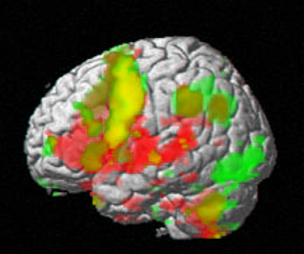
Physicists quickly realized that nuclear magnetic resonance methods had many applications. Relaxation of aligned nuclei, for example, provides a sensitive technique to measure the interaction of nuclei with their own electrons and with other nearby atoms. Imaging came decades later: When a patient is placed in a magnetic field whose strength varies with position, the resonant frequency varies as well, so that RF emissions from relaxing nuclei reveal the positions of atoms. Stronger emission at a certain frequency implies denser tissue at the corresponding location.
Although Rabi’s work was crucial, the MIT and Stanford experiments were “a very big leap,” says Daniel Raftery of Purdue University in Lafayette, Indiana. “The experimental technique was very difficult.” Following Rabi’s 1944 physics Nobel Prize for discovering NMR, Purcell and Bloch shared the 1952 physics Nobel.
–David Lindley
David Lindley is a freelance science writer in Alexandria, Virginia.
References
- F. Bloch, “Nuclear Induction,” Phys. Rev. 70, 460 (1946); F. Bloch, W.W. Hansen, and M. Packard, “The Nuclear Induction Experiment,” Phys. Rev. 70, 474 (1946)
More Information
Nobel Prize info for Bloch and Purcell and for Rabi