Eye Cells as Light Pipes
Computer simulations suggest that a set of cells in the eye could be guiding light, like so many optical fibers, through several layers of cells that cover the light sensitive area at the rear. The team used the optical properties of real cells and a basic equation of wave optics to arrive at the prediction, which they report in the 16 April Physical Review Letters. If the cells’ predicted ability to guide light turns out to be important in vision–still a controversial claim–the result could help explain a bizarre aspect of the eye’s construction.
The retina is at the rear of the eye and includes the light-sensing cells called photoreceptors. But across most of the retina, the photoreceptors are obscured behind three or four coats of additional retinal cells–networked neurons–and a carpet of cellular cables to the brain. Apparently, the retina processes an image by blurring it first. Biologists reference this odd “design” to illustrate that nature’s creations are not all so “intelligent.” Vision scientists have just tried to make sense of how it works as well as it does.
In 2007 a German team reported a study of retinas and cells known as the Müller glia, which stretch from the photoreceptors to the cable layer at the retinal surface [1]. Under the microscope, the team saw the retinas covered with spots where light transmitted through the sample especially easily. Under further investigation, the spots appeared to be single Müller cells acting as optical fibers, or “waveguides.” An array of guides could deliver images to the receptors in-focus and unblurred.
Erez Ribak and his graduate student Amichai Labin of the Technion–Israel Institute of Technology–performed computer simulations to investigate the capacity of Müller cells to guide light. They drew on published measurements of the glial cells’ size, refractive indices, and proximity to one another, as well as properties of cells and fibers abutting them. They calculated the behavior of light illuminating a model cell over a small range of angles, which corresponded to different pupil opening sizes.
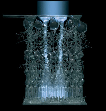
The results were 3D pictures of the amplitude of light waves in and around the illuminated cell. The most common showed waves confined to the cell extending end-to-end in interference patterns known as “guide modes.” Ordinarily, structures of this size tend to scatter visible light, but with guide modes, which are akin to the fundamental and harmonic frequencies in a resonating organ pipe, the cells are transmitting light as the pipe transmits sound waves. So the observation of these modes implies Müller cells can indeed confine and direct light that would otherwise be misdirected by the cells obscuring the photoreceptors.
Brian Vohnsen of University College Dublin says the results are the first to numerically confirm that Müller cells can act as waveguides. It remains to be shown how much of the incident light they guide and how much the photoreceptors depend on them, he says. On the other hand, Vohnsen says, their contribution appears to be where it counts. “They are more or less centered on top of the cone photoreceptors.”
–Oliver Baker
Oliver Baker is a freelance science writer based in Davis, CA.
References
- K. Franze et al., “Müller Cells are Living Optical Fibers in the Vertebrate Retina,” Proc. Natl. Acad. Sci. 104, 8287 (2007)
More Information
Ribak’s web page describing this work, including more videos