Fluid Dynamics Model for Cancer Patterns
The majority of skin cancer deaths arise from melanomas, but this type of tumor is easily treated if detected early. To figure out if a skin tumor is malignant, doctors look for skin discoloration patterns, such as stripes and blobs. However, it’s not clear why these patterns arise or how they depend on tumor type and location. Now Shigeyuki Komura of Tokyo Metropolitan University in Japan and colleagues have developed a numerical model of skin cancer proliferation that reveals a connection between a cancer’s pattern and the underlying fluid dynamics of the skin tissue.
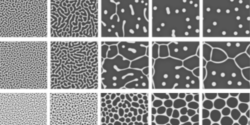
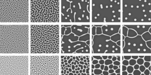
The team’s simulations model the top layer of skin—the epidermis—as a patchwork of healthy and cancerous cells that behave like a 2D fluid. This fluid sits atop a flat surface that represents the dermis, an adjacent skin layer. The researchers alter the strength of the cells’ hydrodynamic interactions—defined as the ratio between the viscosity of the epidermis and the friction between the two skin layers—as well as the cancer cells’ proliferation rate. They then watch to see what patterns arise as the cancer cells reproduce and invade their healthy neighbors.
Some of the resulting patterns match those seen in real-world melanomas. Isolated islands of cancer—a pattern found in soft skin tissues—appear for strong hydrodynamic interactions. And stripes, which are typically observed in stiff skin tissues, such as those found on palms and soles, emerge when hydrodynamic interactions are absent.
The team says that this model cannot yet be used for clinical diagnoses of melanomas. But they hope to develop objective indicators for recognizing the early emergence of these patterns, which may help improve the long-term survival rate for this disease.
This research is published in Physical Review E.
–Christopher Crockett
Christopher Crockett is a freelance writer based in Arlington, Virginia.


