Getting More out of Electron Microscopy
With transmission electron microscopy (TEM), researchers can visualize the atomic structure within a material. Conventional TEM methods, however, often lose valuable phase information that could provide an even more detailed picture of the sample. Now researchers have demonstrated a TEM technique that can recover all available sample information at atomic resolution. The technique could offer researchers a chance to study the electrostatic potential around defects, for example.
Achieving atomic resolution with TEM is challenging, as deflections (aberrations) in the electron paths can distort the images. The sample information can be recovered by using so-called forward modeling, which involves numerically simulating TEM data. Up until now, these simulations have only been achieved for conventional TEM images, in which valuable phase information is missing.
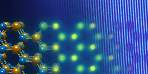
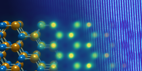
To exploit all of the TEM information, Florian Winkler from the Jülich Research Center in Germany and colleagues devised an automated forward-modeling technique for off-axis electron holography, in which the microscope’s electron beam is split and then recombined to produce an interference pattern. The team’s technique compares holography data to simulations that account for both material properties and microscope characteristics. The simulation’s parameters are then adjusted to give the best fit to the observations. Because the fit is determined directly from the images, the protocol avoids previous problems stemming from time-varying microscope characteristics. As a demonstration, the team performed electron holography on a tungsten diselenide sample and showed that their technique could pick out a small bending of the crystal structure, as well as spatial variations in the aberrations across the image.
This research is published in Physical Review Letters.
–Michael Schirber
Michael Schirber is a Corresponding Editor for Physics based in Lyon, France.


