Silicon Nanowires Feel the Force of Magnetic Resonance
The technique of magnetic resonance force microscopy (MRFM) was conceived as a way to combine the 3D capabilities of magnetic resonance imaging (MRI) with the angstrom-scale resolution of scanning probe techniques, with the ultimate goal of imaging individual biological molecules in three dimensions [1]. The technique makes use of a sensitive mechanical oscillator to detect the minute magnetic forces associated with a small number of magnetic moments, or spins. Compared to conventional magnetic resonance detection methods that use inductive coils to sense nuclear spins, MRFM is about million times more sensitive [2]. With such a dramatic improvement, it is now possible to image virus particles in 3D with nanometer (nm) resolution, far beyond the millimeter resolution of medical MRI. Still, reaching the ultimate goal of single-molecule imaging will require innovations that radically enhance the technique’s sensitivity.
The basic idea behind MRFM is to sense the nuclear spins in a sample by measuring the force that a magnetic field gradient exerts on these moments. Usually, this is accomplished by having the magnetic force drive a small mechanical oscillator, such as a microfabricated cantilever. In one scheme, the sample is attached directly to the cantilever, and the incorporated spins, typically hydrogen nuclei, feel the force from a nearby gradient source. For nanoscale samples, the force can be as small as a few attonewtons ( attonewton = newtons). The key to extending the sensitivity and resolution of MRFM is finding better force transducers to detect these small forces. In an experimental tour de force, John Nichol at the University of Illinois, Urbana-Champaign, and his colleagues report in Physical Review B a significant step in this direction. They have created an oscillator made from a silicon nanowire and used it to detect the magnetic forces on a nanoscale sample of spins [3].
The trick to making a sensitive, low-noise force transducer is to design an oscillator with low stiffness, high resonance frequency, and minimal damping. These requirements inexorably imply low mass, which naturally points to nanometer-scale mechanical devices. The field of nanomechanics has seen explosive growth, with innovative new techniques for creating oscillators from nanotubes, nanowires, and graphene sheets [4]. Nichol et al. have taken advantage of such “bottom-up” fabrication methods to make a high-quality oscillator from a silicon nanowire that is micrometers long and tapers down to a tip with a diameter of less than nm.
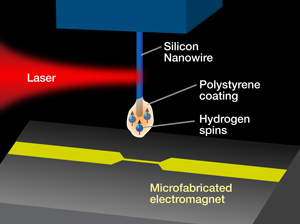
In their demonstration experiment, they coat the tip of the nanowire with hydrogen-rich polystyrene (Fig. 1). The nanowire dangles vertically from a substrate and acts as a cantilever that vibrates in response to oscillating forces applied perpendicular to its length. Since the magnetic forces acting on the hydrogen nuclear spins are so small, the amplitude of the nanowire vibration is only in the angstrom range. Detecting this motion is a major challenge, since the nanowire is so tiny. Nichol et al. have therefore built a special optical interferometer with light polarized along the nanowire axis for enhanced reflectivity. This allows them to achieve the necessary detection sensitivity with minimal heating of the nanowire [3,5]. To generate the magnetic force, they bring the coated tip close to a source of magnetic field gradient, which in their case is produced by running current through a tiny electromagnet consisting of a microfabricated metallic wire. In addition to generating a controllable field gradient, the wire also serves to generate a radio-frequency magnetic field to stimulate magnetic resonance of the hydrogen nuclei.
Achieving attonewton force sensitivity requires the silicon cantilever to have a high mechanical quality factor, . One of the fundamental challenges to force detection, and a significant impediment to further progress in MRFM, is that the factor of a cantilever invariably decreases when its tip is brought close to a surface. This effect is often referred to as “noncontact friction,” and is ubiquitous in near-surface force measurements, though the magnitude of the effect can vary [6–8]. One of the remarkable aspects of Nichol et al.’s nanowire is that it experiences a much lower noncontact friction than is typical for conventional silicon cantilevers. Partly, this is a geometric effect: the small diameter of the nanowire at the tip presents a very small cross section to the surface, giving less potential for interaction. The nanowire also operates at kilohertz (kHz), which is a much higher frequency than the lithographically fabricated cantilevers that have been used previously in MRFM. It may be that the forces from the surface that normally contribute to dissipation primarily fluctuate at lower frequencies, and therefore do not couple as efficiently to a high-frequency oscillator. Either way, the resulting total dissipation is a factor of times lower for the nanowire than for microfabricated cantilevers, leading to a remarkable enhancement in force sensitivity.
In order to actually use these high-frequency oscillators to detect magnetic resonance, the team had to develop an entirely new spin-detection protocol. MRFM typically relies on periodically flipping the nuclear spins of the sample at the cantilever resonance frequency to drive the cantilever vibration. This technique works well as long as the spins’ orientation can be controlled coherently, which is the case when the flipping is performed at a rate of roughly kHz or less. Rather than attempt to manipulate the spins at the much higher cantilever resonant frequency of kHz, Nichol et al. instead create a signal by modulating the magnetic field gradient. By modulating the gradient a few hundred Hz off resonance, and flipping the spins at a few hundred Hz, the force, which is given by the product of the gradient and the spin, has a frequency component right at the cantilever resonance, generating a distinct and unambiguous signal.
With the exceptional level of sensitivity afforded by the nanowire, Nichols et al. are able to detect a spin signal of attonewton (rms) with excellent signal-to-noise ratio. This force is more than a million times smaller than typically detected by conventional atomic force microscopy.
It is the inherently nanomechanical nature of the nanowire resonator, along with its remarkably low noncontact friction, that made these results possible. This marriage of nanomechanics and magnetic resonance represents one possible route to continued progress toward the ultimate goal of extending the resolving power of force-detected magnetic resonance imaging to the molecular level. It also provides a compelling demonstration that nanostructures, such as nanowires, which are based on “bottom-up” fabrication techniques, can indeed become useful transducers to enable new measurement capabilities.
References
- J. A. Sidles, J. L. Garbini, K. J. Bruland, D. Rugar, O. Zueger, S. Hoen, and C. S. Yannoni, Rev. Mod. Phys. 67, 249 (1995)
- C. L. Degen, M. Poggio, H. J. Mamin, C. T. Rettner, and D. Rugar, Proc. Natl. Acad. Sci. U.S.A. 106, 1313 (2009)
- J. M. Nichol, E. R. Hemesath, L. J. Lauhon, and R. Budakian, Phys. Rev. B 85, 054414 (2012)
- See references contained in Ref. 3 for examples of this “bottom-up” approach
- J. M. Nichol, E. R. Hemesath, L. J. Lauhon, and R. Budakian, Appl. Phys. Lett. 93, 193110 (2008)
- B. C. Stipe, H. J. Mamin, T. D. Stowe, T. W. Kenny, and D. Rugar, Phys. Rev. Lett. 87, 096801 (2001)
- S. Kuehn, R. F. Loring, and J. A. Marohn, Phys. Rev. Lett. 96, 156103 (2006)
- A. I. Volokitin and B. N. J. Persson, Phys. Rev. Lett. 95, 086104 (2005)


