Electron Pulses Made Faster Than Atomic Motions
When you read the term “electron beam,” what comes to mind? If you are a physicist or have a background in physics, it may be the great J. J. Thomson—discoverer of the electron—followed by a vision of an old television or oscilloscope powered by cathode-ray tubes. Very twentieth-century stuff. If this is your view, a new study by Jared Maxson at the Pegasus radiation facility at the University of California, Los Angeles, and colleagues [1] should help make clear how electron beams are one of the primary enablers of twenty-first-century science. Ultrashort electron-beam pulses—less than 10 fs long in this case—are enabling forms of atomic-level dynamic imaging that were previously restricted to the realm of thought experiments [2].
It is difficult to overemphasize the impact that electron beams have had on scientific developments over the last century, or the impact they are expected to have over the next century. While electron beams are currently out of favor in high-energy physics, because of the move from electron-positron colliders to hadron colliders at CERN, Fermilab, and other laboratories, they are central to other areas of science. For example, when we want to perform detailed examinations of the structure of molecules and materials, electron beams are at the forefront. Transmission electron microscopes and scanning electron microscopes are remarkably efficient instruments for generating and measuring an enormous range of signals that reveal the structure of materials. These signals come from both the elastic and inelastic interaction of electron beams with materials, and modern materials science is unthinkable without these instruments. When operated at cryogenic temperatures, these same instruments enable 3D reconstructions of proteins, viruses, organelles, and even whole cells [3]. Complementary work can also be performed using synchrotron light sources or free-electron lasers [4], which produce x-ray and infrared beams that are themselves generated from pulsed relativistic electron beams circulating in storage rings or linear accelerators. The size of the facilities that host these instruments, and the properties and limitations of the instruments as sources of x-ray and infrared radiation, are largely determined by the properties of such electron beams.
Historically, the primary developmental frontiers in electron microscopy have been improvements in spatial resolution and new forms of imaging contrast. For x-ray facilities, it has been beam brightness, brilliance, and coherence. Recently, however, a new frontier has emerged: time resolution. This new frontier is inspired by the goal of developing electron and x-ray instruments that can follow the dynamic structure of matter on all relevant spatiotemporal scales. For many chemical reactions, phase transitions, and elementary excitations, this stated goal requires that sub-angstrom atomic displacements be resolved on time scales below 100 fs. That is, it requires time resolution sufficient to follow even the fastest atomic motion while simultaneously maintaining structural sensitivity equivalent to the best conventional approaches. There are two research directions in particular that take advantage of these capabilities and have generated enormous excitement. One is making “molecular movies” that can follow the atomic reorganization of molecules and materials during structural transformations [2]. Another is obtaining atomic images of single particles far beyond the sample’s damage threshold [5]. This latter direction goes by the name “diffract and destroy.”
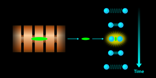
Against this backdrop of interest in ultrashort electron pulses, Maxson and colleagues’ work at the Pegasus facility is a landmark. The authors have demonstrated, using an electron source similar to that used to fill the storage ring at synchrotron facilities, that a very high quality electron beam with pulses of duration below 10 fs can be produced. For perspective, the highest-frequency phonons (lattice vibrations) in diamond—the hardest natural material—have a period of approximately 20 fs. Only the vibrations of molecules involving hydrogen atoms (C-H, O-H, and N-H) have periods as short as about 10 fs. That is, on time scales below 10 fs, essentially all atomic motion is frozen in molecules and materials. A snapshot of structure taken with 10-fs pulses would represent a truly instantaneous atomic configuration (Fig. 1). The researchers were able to obtain such short pulses by exploring an operating regime of the source that is not particularly attractive for use at synchrotrons and x-ray free-electron-laser facilities (the usual application areas for such electron sources), but that produces beams with very attractive properties for direct use in dynamic-imaging experiments in terms of pulse duration and beam size. The payoff is unprecedented time resolution with a beam diameter capable of probing individual micrometer-sized crystals. The result opens up completely new scientific territory: microcrystallography with 10-fs time resolution. Conventional crystallography makes use of x-ray or electron diffraction patterns to determine atomic structure in equilibrium. With electron pulses as short as 10 fs, researchers will be able to explore the far-from-equilibrium atomic structure of molecules and materials in exquisite detail. That is, we will be able to follow the time-evolving atomic structure with no “blurring” of the motions. The micrometer-sized beams mean that millimeter (or larger) crystals, often difficult to produce, will not be necessary for such studies.
While the Pegasus facility is currently the only one of its kind on a university campus in North America (all other similar facilities are to be found at national labs [6]), these results may lead to more such facilities in the near future. Recently, there has been something of an explosion of interest in methods to produce ultrashort electron-beam pulses in university labs around the world. This is due to the significant scientific impact these approaches are having in materials and molecular physics [7–10], and the relative accessibility of the technology in terms of cost. Overwhelmingly, these new labs are based on commercial transmission-electron-microscopy platforms [10] or homebuilt instrumentation that also operate at beam energies of 50–300 keV [11], rather than the MeV energies used in the Pegasus instrument and similar devices. Some of these instruments even use devices to compress the electron pulses in a manner similar to the current work [12].
The higher beam energy of Pegasus carries with it significant additional cost, complexity, and even radiation safety issues. But it also has several important fundamental advantages. The impact of Coulomb repulsion inside the ultrashort electron pulses that determines instrument capabilities and performance (such as pulse duration and fluence) at the nonrelativistic beam energies is dramatically reduced through relativistic effects. This factor helps to explain why both the electron-pulse duration (10 fs) and the beam diameter (5 micrometers) demonstrated in the current work are both roughly an order of magnitude smaller than the state of the art in nonrelativistic instruments at the same number of electrons per pulse. A factor of 10 in both of these key metrics means that qualitatively new types of measurements will be possible. The next step is thus to start thinking up great measurements that could take advantage of this breakthrough.
This research is published in Physical Review Letters.
References
- J. Maxson et al., “Direct Measurement of Sub-10 fs Relativistic Electron Beams with Ultralow Emittance,” Phys. Rev. Lett. 118, 154802 (2017).
- G. Sciaini and R. J. D. Miller, “Femtosecond Electron Diffraction: Heralding the Era of Atomically Resolved Dynamics,” Rep. Prog. Phys. 74, 096101 (2011).
- R. Fernandez-Leiro and S. H. W. Scheres, “Unravelling Biological Macromolecules with Cryo-Electron Microscopy,” Nature 537, 339 (2016).
- C. Pellegrini, A. Marinelli, and S. Reiche, “The Physics of X-Ray Free-Electron Lasers,” Rev. Mod. Phys. 88, 015006 (2016).
- J. Spence, “X-Ray Imaging: Ultrafast Diffract-And-Destroy Movies,” Nature Photon. 2, 390 (2008).
- P. Zhu et al., “Femtosecond Time-Resolved MeV Electron Diffraction,” New J. Phys 17, 063004 (2015).
- T. Ishikawa et al., “Direct Observation of Collective Modes Coupled to Molecular Orbital–Driven Charge Transfer,” Science 350, 1501 (2015).
- V. R. Morrison, R. P. Chatelain, K. L. Tiwari, A. Hendaoui, A. Bruhacs, M. Chaker, and B. J. Siwick, “A Photoinduced Metal-Like Phase of Monoclinic VO2 Revealed by Ultrafast Electron Diffraction,” Science 346, 445 (2014).
- L. Waldecker, T. A. Miller, M. Rudé, R. Bertoni, J. Osmond, V. Pruneri, R. E. Simpson, R. Ernstorfer, and S. Wall, “Time-Domain Separation of Optical Properties from Structural Transitions in Resonantly Bonded Materials,” Nature Mater. 14, 991 (2015).
- D. J. Flannigan and A. H. Zewail, “4D Electron Microscopy: Principles and Applications,” Acc. Chem. Res. 45, 1828 (2012).
- C. Gerbig, A. Senftleben, S. Morgenstern, C. Sarpe, and T. Baumert, “Spatio-Temporal Resolution Studies on a Highly Compact Ultrafast Electron Diffractometer,” New J. Phys. 17, 043050 (2015).
- R. P. Chatelain, V. R. Morrison, C. Godbout, and B. J. Siwick, “Ultrafast Electron Diffraction with Radio-Frequency Compressed Electron Pulses,” Appl. Phys. Lett. 101, 081901 (2012).


