Using Physics to Speed up Tissue Engineering
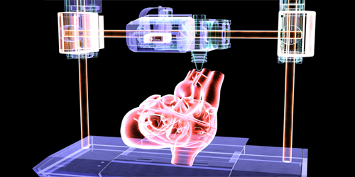
Bioprinting—the 3D printing of human tissues—holds potential for producing tissues and organs for transplants. In this technology, a machine dispenses small segments of bioinks—materials made of cell aggregates or microtissues. When placed next to each other, the segments fuse and self-assemble into sturdier tissue structures. However, because the self-assembly process is slow, tissues assembled using a patient’s own cells cannot be printed in urgent clinical settings. Guided by a physics-based microscopic model, Ashkan Shafiee of Wake Forest School of Medicine, North Carolina, and colleagues have devised a new bioprinting method that accelerates tissue self-assembly [1].
Their method, based on bioink fusion, is an iterative process in which cells in one printed microtissue break off to develop new chemical bonds with an adjacent tissue. From a macroscopic perspective, this microscopic bond formation resembles a closing zipper. The researchers designed their method by harnessing a model prediction—that the bioinks would fuse faster when subjected to an external force. The model allowed them to optimize the magnitude of the force to accelerate bioink fusion without damaging existing cells.
In experiments, the team created a force with the desired magnitude by printing the bioinks into semicircular-shaped grooves on a gel platform. The grooves’ geometry produced a rotational torque and other mechanical forces that caused the bioink segments to press against each other. By imaging the structural evolution of the fusing bioinks, they found that fusion occurred 3 times faster in the grooves than it did on a flat platform. The researchers are currently developing a bioprinter in which the forces on the bioinks can be finely tuned.
–Sophia Chen
Sophia Chen is a freelance science writer based in Columbus, Ohio.
References
- A. Shafiee et al., “Acceleration of tissue maturation by mechanotransduction-based bioprinting,” Phys. Rev. Research 3, 013008 (2021).