Making Miniature Artificial Cilia
The single-celled paramecium whizzes through its aqueous habitat at a rate of 10 times its length a second, thanks to the coordinated undulation of hair-like structures, called cilia. Cilia are also found in human lungs where their motion helps to clear out mucus. Researchers in microfluidics would like to mimic cilia’s fluid-shifting ability for novel devices, but they have had trouble creating artificial cilia at the microscale. Now Jaap den Toonder of Eindhoven University of Technology in the Netherlands and his collaborators have demonstrated miniaturized artificial cilia that perform like real ones [1]. What’s more, their experiments clarified which aspects of the cilia’s motion are responsible for fluid shifting.
Natural cilia consist of bundles of microtubules and other specialized proteins. Their coordinated flexing is orchestrated by a bucket brigade of signaling molecules that triggers a given cilium to flex moments after its neighbor does. The resulting pattern, called metachrony, resembles a crowd wave in a sports stadium. Researchers can reproduce this collective motion in artificial cilia with the help of a time-varying magnetic field. In this scheme, the cilia are flexible rods made of a paramagnetic material and arranged as an array on a surface. The field sweeps across the array, like an airport radar scanning across the sky, and the cilia respond by bending in the direction of the field. Den Toonder and his team realized this concept two years ago using rods activated by a row of magnets on a conveyor belt. But the rods and magnets were around 10 mm in size—3 orders of magnitude larger than natural cilia.
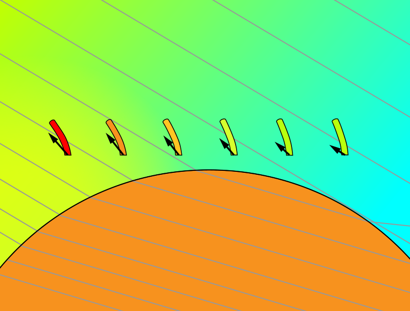
The team’s new experiment manages to shrink the size by 2 orders of magnitude, and there’s a possibility to go even smaller. What made the miniaturization of the system possible was the new way in which the magnetic field controlled the artificial cilia. The researchers fabricated their 150-µm-long cilia from paramagnetic carbonyl iron using standard techniques. They then attached arrays of these cilia to a plastic substrate, which sat above a cylinder of paramagnetic material. When a rotating magnetic field was applied to the system, the cylinder distorted the field lines in such a way that each cilium felt a different magnetic field at any given time. As this local field cycled from strong to weak, each cilium responded by bending forward and then snapping back elastically. The timing of the field variations was tuned—using simulation software—so that a wave traveled through the array.
Using a video camera attached to a microscope, den Toonder and his collaborators verified that their miniaturized artificial cilia exhibited metachronal motion. But could the cilia shift fluid? And if so, how? The question is subtler than it might seem. Conceivably, the whip-like motion of individual cilia and their collective metachronal wave could both drive fluid flow. What’s more, if an individual cilium’s forward and backward strokes are symmetric, the cilium can’t produce net fluid motion by itself.
To find answers, the team made a microfluidic device out of transparent plastic and filled it in turn with two liquids: water to represent a low-viscosity fluid and glycerol to represent a high-viscosity fluid. They also explored two types of cilia arrays—one in which the wave propagated in the same direction as the forward-bending direction and another in which the wave and bending directions were opposite. These two cases are respectively called symplectic and antiplectic, and they correspond to the two main types of metachronal motion observed in natural cilia. Removing the paramagnetic cylinder from the setup but using the same rotating field caused the cilia to oscillate back and forth without generating a metachronal wave.
By analyzing the video, the researchers could track the cilia tip motion for both the forward, magnetically induced stroke and the backward, elastic-restoring stroke. They found that for all the various scenarios the magnetic stroke swept more area than the elastic stroke, which would presumably imply that the magnetic stroke dominates the flow generation. Indeed, for the high-viscosity fluid, the measured fluid flow was in the forward direction. However, in the low-viscosity fluid, the elastic stroke was much faster than the magnetic one, and this whip-like action led to backward flow. In both fluids, the metachronal motion was found to bias the cilia-generated flow, enhancing it for antiplectic metachrony and decreasing it for symplectic metachrony.
The results demonstrate the different ways in which individual and collective cilia motion can contribute to flow generation. The researchers say that this information may benefit future efforts to integrate artificial cilia into small-scale microfluidic chips. They also foresee further miniaturization of artificial cilia to the scale of natural cilia, allowing investigations into how cells and tissues manage to control their fluid environment.
–Charles Day
Charles Day is a Senior Editor for Physics Magazine.
References
- Z. Cui et al., “Miniaturized metachronal magnetic artificial cilia,” Proc. Natl. Acad. Sci. U.S.A. 120 (2023).