




More Than One Way to Assemble Molecular Scaffolding
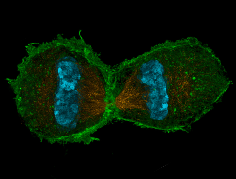

A biomolecular condensate is a small region within a cell where proteins, RNA, or other biomolecules are highly concentrated and perform a specific task, such as accelerating a chemical reaction. Now researchers may have uncovered a previously unknown role for condensates: the bundling of scaffolding proteins called microtubules into thicker, stronger fibers [1]. The researchers used two experimental techniques and theoretical modeling to study mixtures of microtubules and proteins known to associate with them. The results suggest that cells use an additional mechanism besides chemical binding to bundle microtubules.
Microtubules are rigid biopolymers 25 nm in diameter and as much as a few micrometers in length. Among the roles they fulfill is the facilitation of cell division by forming a structure called the mitotic spindle, which includes microtubule fibers along which chromosomes move before a cell divides. The forces involved would break a single microtubule, so spindle fibers are made up of bundles of up to 30 microtubules.
Surface charges cause a strong electrostatic repulsion between microtubules, so bundling requires so-called microtubule-associated proteins (MAPs) to chemically link pairs of microtubules, which starts the bundling process. But MAPs also form biomolecular condensates and do so in the vicinity of microtubule bundles. To biophysicist Sabine Petry of Princeton University, the fact that several different MAPs can condense was a hint that MAPs might be capable of bundling microtubules using capillary forces, in addition to chemical bonds.
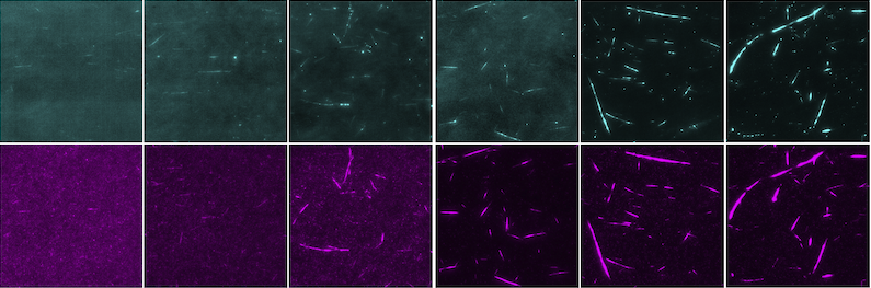
To test her hypothesis, Petry and her colleagues exposed fluorescently tagged microtubules to two fluorescently tagged MAPs, TPX2 and BuGZ. TPX2 has two sites that bind to microtubules, so it can bundle pairs of them through chemical binding, but BuGZ has just one such site.
The researchers used fluorescence microscopy to observe each MAP in a solution of microtubules. In each case, the team triggered the condensation and bundling by gradually increasing the MAP concentration. The condensates stuck to the bundles in clumps, like beads of liquid along a wet thread. The two MAPs behaved identically even though only TPX2 can bundle microtubes via chemical binding alone.
Petry and her colleagues suspected that MAP condensates bundle microtubules by forming capillary bridges—thin, sticky films of condensate. Driven by surface tension, such liquid bridges are responsible for clumping strands of wet hair and for enabling a licked finger to turn the page of a book. The researchers used atomic force microscopy to directly measure the surface tension of the condensates coating the microtubules. The measured values turned out to be large enough to pull microtubules together, which supports the idea that microtubules can be bundled by capillary bridges.
The researchers theoretically analyzed the bundling assuming two different bridging mechanisms: chemical binding via linker molecules and capillary forces via liquid bridges. The process begins with one microtubule lying atop another and the two polymers oriented in different directions and ends with the two aligning and snapping together. The snapping initially proceeds at the same rate for both mechanisms because of the resistance of the solvent to the twisting motion, the analysis indicated. But when the microtubules are nearly aligned, a second source of resistance appears for the capillary mechanism but not for the chemical one: the need to squeeze out the condensate before bundling is complete. Although current techniques lack the resolution to resolve the squeezing, this difference could in principle be used as way to measure and compare the degree to which the two mechanisms are used in cells.
Future experiments may also address whether MAP condensates are involved in bundling in real cells. The counter argument is that such a delicate, choreographed process as cell division might be easily disrupted if it relied on MAPs to suddenly increase in concentration enough to condense. But team member Howard Stone of Princeton points out that cells routinely run out-of-equilibrium processes that push condensation to happen “at the right place and the right time.” Indeed, TPX2 is known to be sequestered at the onset of cell division. Shortly after, a protein bound to the surfaces of chromosomes activates factors that release TPX2, locally concentrating it near chromosomes, where, Stone says, it could condense and bundle microtubules.
“This is a novel observation,” says biologist Scott Forth of Rensselaer Polytechnic Institute in New York. He says that TPX2 and BuGZ are important for maintaining spindle integrity during cell division. “This new bundling property likely has consequences in cell biology but also human health concerns.” A malfunction or malformation of the mitotic spindle is a primary driver of several severe human diseases, including cancer and genetic neurodevelopmental disorders.
–Charles Day
Charles Day is a Senior Editor for Physics Magazine.
References
- B. Gouveia et al., “Capillary bundling of microtubules by condensates,” PRX Life 4, 043006 (2026).