X Rays Expose Transient Spins
The possibility that spin-polarized electrons can travel over relatively long distances in nonmagnetic materials, such as copper, offers exciting prospects for applications in spintronics and information technology [1, 2]. Experimental and theoretical studies have shown that spin-polarized electrons injected from a ferromagnet into a nonmagnetic material give rise to a spin accumulation (magnetization), and a spin current, in the nonmagnetic material. Notwithstanding a large body of experimental work, evidence for such a spin accumulation at the nanoscale has so far been lacking, because it requires sophisticated techniques that can probe directly the magnetic moments induced in the nonmagnetic material. Now, Jo Stöhr and colleagues [3] report how they have used x-ray spectromicroscopy to detect the transient magnetic moments in a nonmagnetic metal layer (copper) caused by spin injection from an adjacent ferromagnetic layer (cobalt).
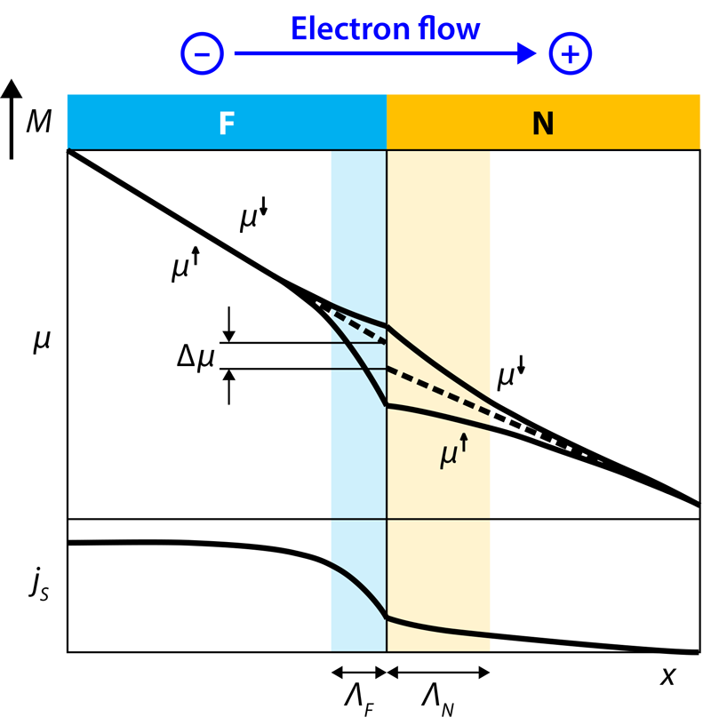
The origin of spin accumulation across a ferromagnet–nonmagnetic material (F/N) interface can be clarified as follows [4]. Assume the ferromagnet has an excess of spin-down electrons over spin-up electrons. When a voltage is applied to the structure so that electrons flow from the F side to the N side, more spin-down electrons travel across the interface. This spin imbalance leads to the accumulation of spin-down electrons near the interface, which diffuse away from it into both sides. The difference in the chemical potentials for each spin direction at each point in the structure determines the spin current: on the F side, the spin current increases with distance from the interface until it reaches a plateau value, whereas on the N side, it decays and eventually vanishes because of spin-flip scattering (Fig. 1).
In their study, Stöhr and colleagues used a multilayer nanopillar structure comprising a ferromagnetic cobalt (Co) layer and a neighboring nonmagnetic copper (Cu) layer. The small dimensions of the nanopillar allow a charge current of high density to flow in the structure, which is necessary to observe the spin accumulation, while any undesired magnetic field produced by this current remains low. The authors obtained high-resolution x-ray absorption spectra of the nanopillar using the scanning transmission x-ray microscope at the Stanford Synchrotron Radiation Lightsource.
To detect the transient magnetic moments in the Cu layer, Stöhr and co-workers obtained an x-ray magnetic circular dichroism (XMCD) spectrum. This spectrum is determined from the difference between two x-ray absorption spectra: one with the circular polarization direction of the x-rays parallel to the magnetization direction of the structure, and the other with the polarization direction antiparallel to it [5]. The spin-polarized core electrons in a magnetized material, excited by x-rays into the unoccupied conduction states, generate a strong magneto-optical effect that is observable in the XMCD spectrum. This XMCD signal can be used to determine the difference in spin-up and spin-down electrons on the material’s excited atoms, and, as a result, their magnetic moments. By switching the charge current flowing through the structure periodically on and off, the authors were able to obtain a high sensitivity to the small XMCD signal associated with the Cu atoms, which allowed them to detect their transient magnetic moments. Moreover, in agreement with the expectation for spin accumulation at an F/N interface, the measurements showed that the XMCD signal reverses in sign with the direction of the applied current as well as with the x-ray polarization direction.
Going into the details, the XMCD spectrum of the Cu layer revealed two distinct peaks as a function of photon energy. The first peak is caused by spin accumulation across the entire Cu layer, and its magnitude was found to correspond to a transient magnetic moment of per Cu atom for an electron current of 5 milliamps; is the Bohr magneton, the unit used for expressing the magnetic moment of an electron. The second, and broader, peak, located at a photon energy that is 0.7 electron volts higher than the first peak, is due to the magnetization of Cu atoms at the interface, which is induced by hybridization (mixing) between Cu and Co electronic states. The authors find that the magnetization at the Cu interface is transiently increased by about 10%, which amounts to a magnetic moment of per Cu atom—about 2 orders of magnitude larger than for the bulk Cu atoms.
Curiously, the measured spin accumulation in the bulk of the Cu layer turns out to be 3 times smaller than expected from modeling spin diffusion across F/N interfaces. Stöhr and colleagues ascribe this deficit in the spin accumulation to the presence of an interfacial spin-torque process. Clearly, more work is required to comprehend this discrepancy, such as measuring the dynamic response of the spin accumulation near the F/N interface. Studying spin torque and “spin pumping” at F/N interfaces and spin valves formed by an F1/N/F2 multilayer, where F1 and F2 layers represent the ferromagnetic spin source and sink respectively, is of great interest for device applications [6].
To understand spin pumping, suppose the magnetization at the F/N interface is switched very fast; this happens if the ferromagnetic layer has a precessing magnetization. In this case, the difference in the chemical potentials of the spin-up and spin-down electrons leads to a large nonequilibrium spin current. This current can be injected into the nonmagnetic material through the effect of ferromagnetic resonance, creating a pure spin source, or “spin battery” [7]. Spin pumping into the nonmagnetic material has been observed indirectly as an increased damping of the ferromagnetic resonance [8]. However, XMCD makes it possible to study the magnetization dynamics of the source and sink layers independently. Important steps in this direction have recently been taken, by evidencing the spin pumping using x-ray ferromagnetic resonance experiments in which the precessing magnetic moments are excited by microwave radiation and probed with XMCD [9, 10].
This research is published in Physical Review Letters.
References
- I. Žutić, J. Fabian, and S. Das Sarma, “Spintronics: Fundamentals and applications,” Rev. Mod. Phys. 76, 323 (2004).
- Spin Current, edited by S. Maekawa, S. O. Valenzuela, E. Saitoh, and T. Kimura (Oxford University Press, Oxford, 2012)[Amazon][WorldCat].
- R. Kukreja, S. Bonetti, Z. Chen, D. Backes, Y. Acremann, J. Katine, A. D. Kent, H. A. Dürr, H. Ohldag, and J. Stöhr, “X-Ray Detection of Transient Magnetic Moments Induced by a Spin Current in Cu,” Phys. Rev. Lett. 115, 096601 (2015).
- M. D. Stiles and J. Miltat, “Spin Transfer Torque and Dynamics,” Top. Appl. Phys. 101, 225 (2006).
- G. van der Laan and A. I. Figueroa, “X-ray Magnetic Circular Dichroism—A Versatile Tool to Study Magnetism,” Coord. Chem. Rev. 277-278, 95 (2014).
- Y. Tserkovnyak, A. Brataas, G. E. W. Bauer, and B. I. Halperin, “Nonlocal Magnetization Dynamics in Ferromagnetic Heterostructures,” Rev. Mod. Phys. 77, 1375 (2005).
- A. Brataas, Y. Tserkovnyak, G. E. W. Bauer, and I. Halperin, “Spin Battery Operated by Ferromagnetic Resonance,” Phys. Rev. B 66, 060404 (2002).
- B. Heinrich, Y. Tserkovnyak, G. Woltersdorf, A. Brataas, R. Urban, and G. E. W. Bauer, “Dynamic Exchange Coupling in Magnetic Bilayers,” Phys. Rev. Lett. 90, 187601 (2003).
- M. K. Marcham et al., “Phase-Resolved X-Ray Ferromagnetic Resonance Measurements of Spin Pumping in Spin Valve Structures,” Phys. Rev. B 87, 180403 (2013).
- J. Li et al., “Direct Detection of Pure Spin-Current by X-Ray Pump-Probe Measurements,” arXiv:1505.03959 .


