Better than Diffraction
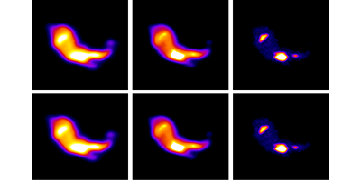
Optical microscopes are widely used in all areas of science to image small objects. However, because of their design and the limits of diffraction, the smallest features that conventional microscopes can image are about half the wavelength of the light they use. Writing in Physical Review Letters, Shi-Wei Chu, at the National Taiwan University, and colleagues report a new technique that overcomes this resolution limit and can image nanostructures 70 nanometers in size—less than one eighth of the wavelength of the visible light used in their setup.
The group fitted a standard optical microscope with a laser and used it to image a sample containing gold nanoparticles. The laser wavelength was chosen so that it was resonant with the sharp plasmonic resonance exhibited by the particles. As a consequence, the laser light experienced particularly strong scattering. By adjusting the intensity of the laser, the researchers were able to reach, for the first time, a regime in which the light scattered from an isolated particle was saturated. With proper image processing techniques, such saturation behavior could be exploited to deliver sharper images of the plasmonic nanostructures.
While this method only works for gold nanoparticles, the particles can be selectively embedded into other materials, allowing them to be imaged. Although other recently demonstrated techniques, mostly based on fluorescence microscopy, allow comparable or even better resolution, this gold-nanoparticle method has an important advantage: samples can be imaged repeatedly without damage and with no loss in scattering intensity that, in fluorescence-based schemes, inevitably occur because of the bleaching of the fluorescing molecules. – Katherine Wright