Brain Tissue Amplifies Waves
Few experiments have been able to visualize the brain’s response to a blow to the head. A new imaging study analyzes wave-like disturbances in the brain that might be generated from a head impact. The researchers find that so-called shear waves, which push on a medium perpendicular to their direction of travel, amplify the acceleration experienced by neurons as the waves move through the brain. The finding suggests the waves may play an important role in head-trauma injuries.
Brain tissue is a nonlinear material, meaning that waves traveling through it are likely to become distorted. But whether these effects matter in head trauma has been difficult to assess. That’s because most imaging techniques lack the necessary combination of spatial resolution and penetration depth.
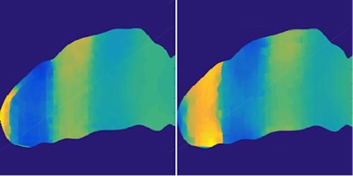
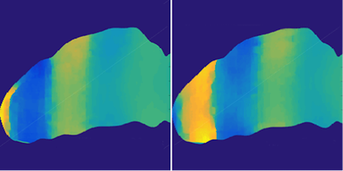
To acquire their images, Gianmarco Pinton and colleagues at the University of North Carolina, Chapel Hill, developed an ultrasound technique for monitoring tissue distortions in response to shear waves at high speed and with high resolution. The researchers immerse a pig brain in gelatin, in which they jiggle a plate up and down to produce shear waves that penetrate the brain. The team captured the wave profiles at about 6000 frames per second and with a resolution of 1 m, or a tenth the width of a blood cell. This is enough spatial and time resolution to see that, after entering the brain, the initially smooth waves quickly develop a shock front that provides 9 times more acceleration to the surrounding tissue. It is, however, an open question whether these findings apply to real head traumas, in which the waves would have to pass from the skull through the meninges and cerebrospinal fluid.
This research is published in Physical Review Applied.
–Jessica Thomas
Jessica Thomas is the Editor of Physics.


