Positron Emission Tomography Could Be Aided by Entanglement
Medical scans using an imaging method called positron emission tomography (PET) are crucial for diagnosing diseases such as cancer and Alzheimer’s. In PET, electrons and positrons annihilate into pairs of photons inside a patient’s body, and the photons are detected and used to reconstruct images of body tissues. The photons in each pair are known to be quantum entangled in their polarization, and recent work has suggested that this entanglement could improve the quality of PET imaging. Such quantum-enhanced imaging could now be one step closer thanks to Julien Bordes and colleagues at the University of York, UK, who have observed that the photon entanglement is much more resilient than previously thought [1].
Currently, in PET, a patient receives an intravenous injection of biomolecules with attached radioactive atoms that emit a positron as they decay. The annihilation of such a positron with an electron in the patient’s body creates two photons that propagate in opposite directions, each with an energy of 511 keV—more than 100,000 times the energy of visible light. Such photons can penetrate through the patient’s body and produce signals in the PET detectors. These signals enable the determination of the distribution of electron–positron annihilations in the body and, in turn, the production of images showing how fast the administered biomolecules are metabolized in body tissues.
Over the past 70 years, PET has undergone continuous development, moving from blurred images obtained using only two detectors to dynamic, simultaneous full-body imaging obtained using state-of-the-art systems constructed from hundreds of thousands of crystal scintillators. However, the main principle of PET scanners has remained unchanged: They reconstruct the distribution of electron–positron annihilations by recording when and where the created photons interact with the detector system and how much energy they deposit.
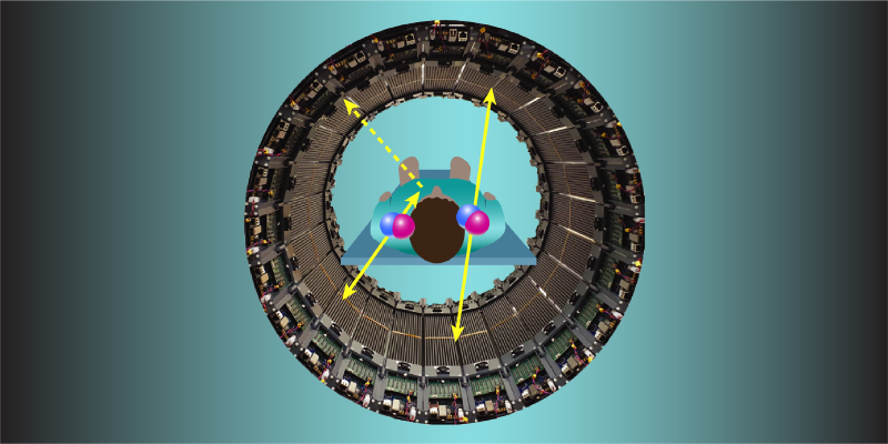
A key challenge in PET imaging is filtering out events in which one or both photons from an annihilation scatter off an electron in the patient’s body before reaching the detectors (Fig. 1). Such filtering is crucial because these events account for about 90% of all detected photon pairs and cause PET images to be blurry. The scattered photons have less energy than the original photons, so some of the blurring events can be filtered out by rejecting photons with measured energies lower than 511 keV (by a margin greater than the detector’s energy resolution).
In 2014, scientists proposed that blurring events could be suppressed by carefully examining the difference in the polarization direction of two photons coming from the same annihilation [2]. This method works only if, after one photon scatters in the patient’s body, the photons do not remain entangled in their polarization and begin to propagate independently of each other [3]. Until a few years ago, this entanglement loss was widely thought to occur [4]. But in 2023, an experiment indicated that entanglement persisted after one photon from an entangled pair scattered [5]. This unexpected observation was confirmed by independent experiments for scattering angles up to 50°, with a hint of entanglement loss noted only at 50° [6].
Bordes and colleagues extended these studies to a scattering angle of 70°, which allowed them to observe the first clear evidence for entanglement loss at angles larger than 50°. Moreover, by considering the scattering of one of the two entangled photons as well as entanglement-loss effects, the team showed that the dependence of the degree of entanglement on the scattering angle is well described by a recently developed quantum theory [7]. This theory assumes the probability that a photon from an entangled pair scatters off an electron at a given angle depends not only on the photon’s momentum and polarization but also on the degree of entanglement between the photons.
The observation that the photons created in an electron–positron annihilation can remain entangled when one of them is scattered is a scientifically exciting discovery, but it is both bad and good news for medical diagnosis. It is bad news because it means that measuring the difference between photon polarizations cannot help in PET imaging by reducing the fraction of blurring events caused by scattering in the patient’s body. But it is potentially good news for the development of quantum-enhanced PET diagnosis because the possible diagnostic information about body tissues that is carried by entangled photons will not be lost if one photon scatters in the body.
The first full-scale PET system capable of entanglement-aided imaging has already been constructed using plastic scintillators [8]. Preliminary results with this system, known as J-PET, have demonstrated a dependence of the degree of entanglement on the type of material in which the electron–positron annihilations occur—a promising indication of the potential use of entanglement in PET diagnosis. Additionally, several groups are working on the development of technology for conventional, crystal-scintillator-based PET systems capable of entanglement-aided imaging [1, 4, 9, 10].
In this decade, the advancement of PET is undergoing a paradigm shift toward completely new diagnostic parameters. Such parameters might be sensitive to how the electron–positron annihilations occur, given the dependence of the lifetime of positronium (a bound electron–positron state) and the degree of entanglement on the type of tissue [8]. The intriguing results on photon entanglement reported by Bordes and colleagues could act as an invitation to the broader scientific community to join this emerging field.
References
- J. Bordes et al., “First detailed study of the quantum decoherence of entangled gamma photons,” Phys. Rev. Lett. 133, 132502 (2024).
- A. L. McNamara et al., “Towards optimal imaging with PET: An in silico feasibility study,” Phys. Med. Biol. 59, 7587 (2014).
- P. Moskal et al., “Feasibility studies of the polarization of photons beyond the optical wavelength regime with the J-PET detector,” Eur. Phys. J. C 78, 970 (2018).
- D. P. Watts et al., “Photon quantum entanglement in the MeV regime and its application in PET imaging,” Nat. Commun. 12, 2646 (2021).
- A. Ivashkin et al., “Testing entanglement of annihilation photons,” Sci. Rep. 13, 7559 (2023).
- S. Parashari et al., “Closing the door on the ‘puzzle of decoherence’ of annihilation quanta,” Phys. Lett. B 852, 138628 (2024).
- P. Caradonna, “Kinematic analysis of multiple Compton scattering in quantum-entangled two-photon systems,” Ann. Phys. 470, 169779 (2024).
- P. Moskal et al., “Non-maximal entanglement of photons from positron-electron annihilation demonstrated using a novel plastic PET scanner,” arXiv: 2407.08574; “Positronium image of the human brain in vivo,” Sci. Adv. 10, eadp2840 (2024).
- G. Romanchek et al., “Application of quantum entanglement induced polarization for dual-positron and prompt gamma imaging,” Bio-Algorithms Med-Syst. 19, 9 (2023).
- D. Kim et al., “Background reduction in PET by double Compton scattering of quantum entangled annihilation photons,” J. Instrum. 18, P07007 (2023).